
Intranodular hyperechogenic figures - case 443 (cytologic picture 1)
|
|

|

|
|
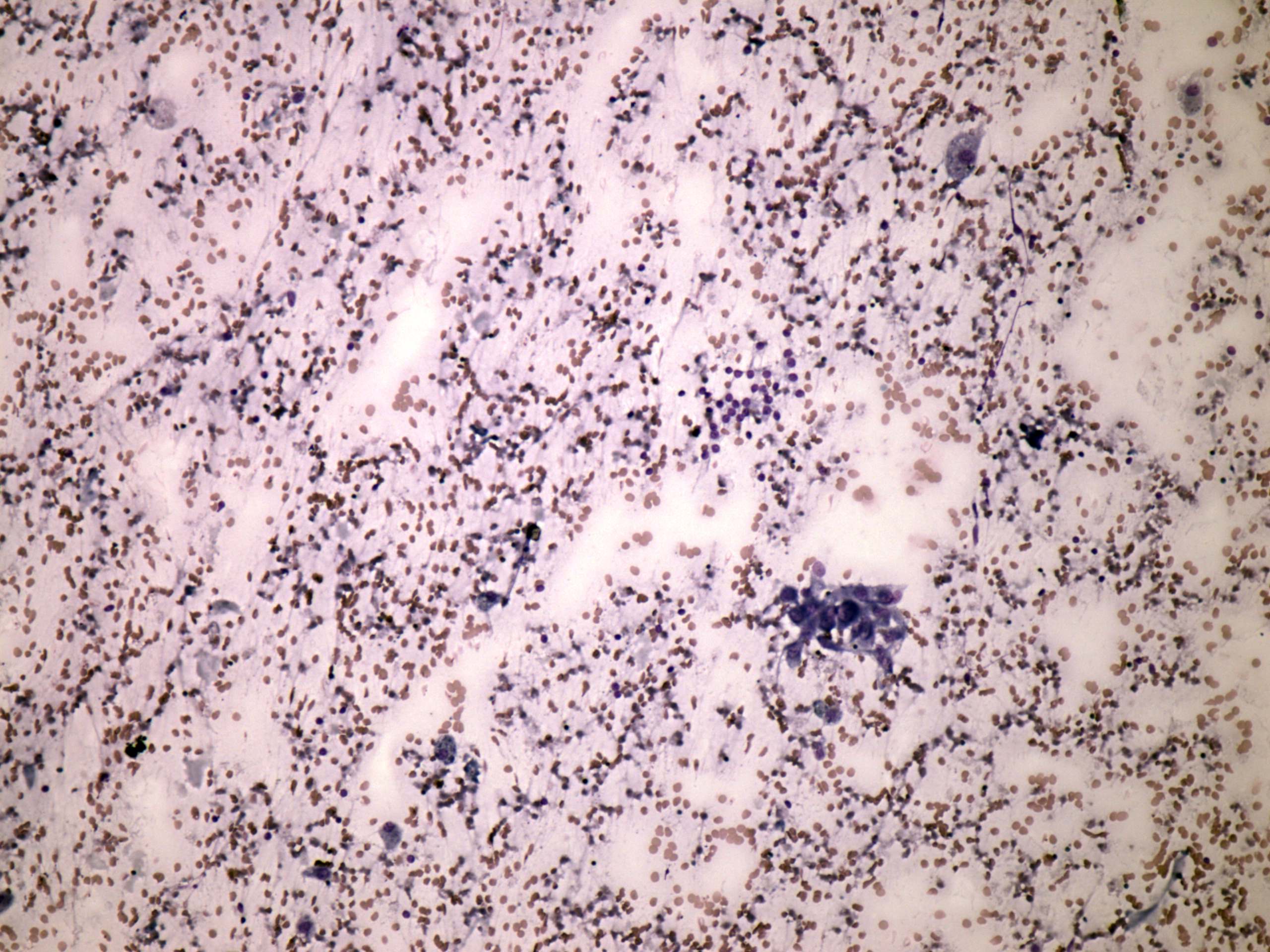
Wright-Giemsa staining, 100x. There is a small, regular monolayered fragment composed of typical follicular cells and there are numerous macrophages.