
Intranodular hyperechogenic figures - case 662 (ultrasonographic picture 2)

|

|

|
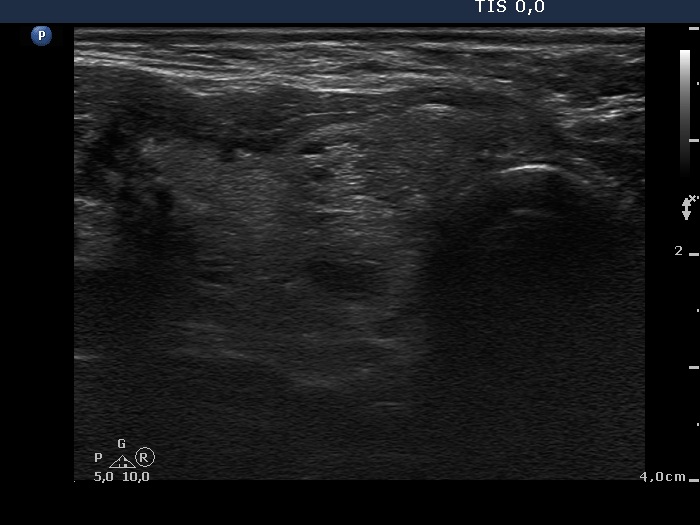
Lower part of the right lobe, another transverse view. There is a hyperechogenic nodule in the lower part of the lobe. This lesion presents numerous back wall cystic figures in the form of echogenic lines and granules.


