
|
|
Intranodular hyperechogenic figures - case 694
|
|


Clinical data: A 36-year-old woman came to a routine follow-up. She has been known having hypothyroidism and nodular goiter for 12 years.
Palpation: a moderately firm nodule in the isthmic part of the left lobe.
Hormonal evaluation: TSH 5.28 mIU/L on daily 125 microgram levothyroxine.
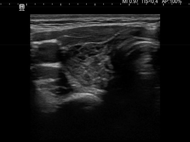
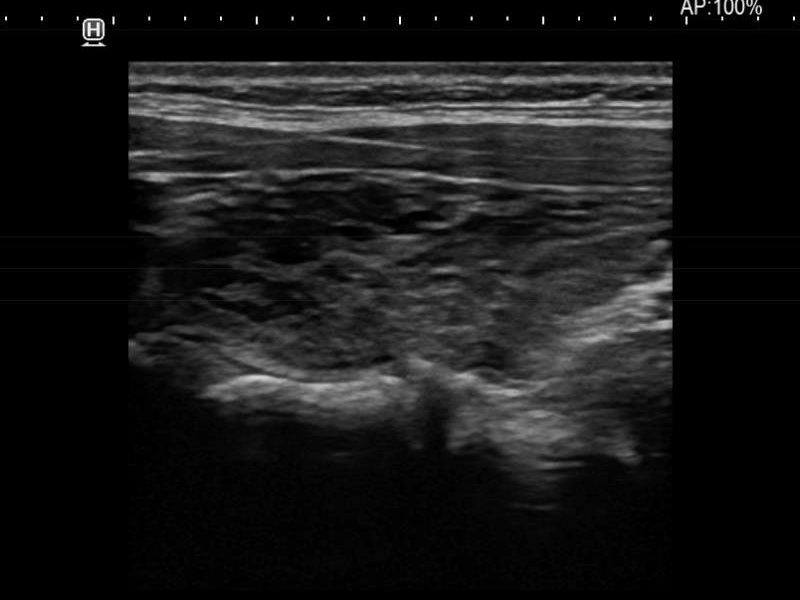
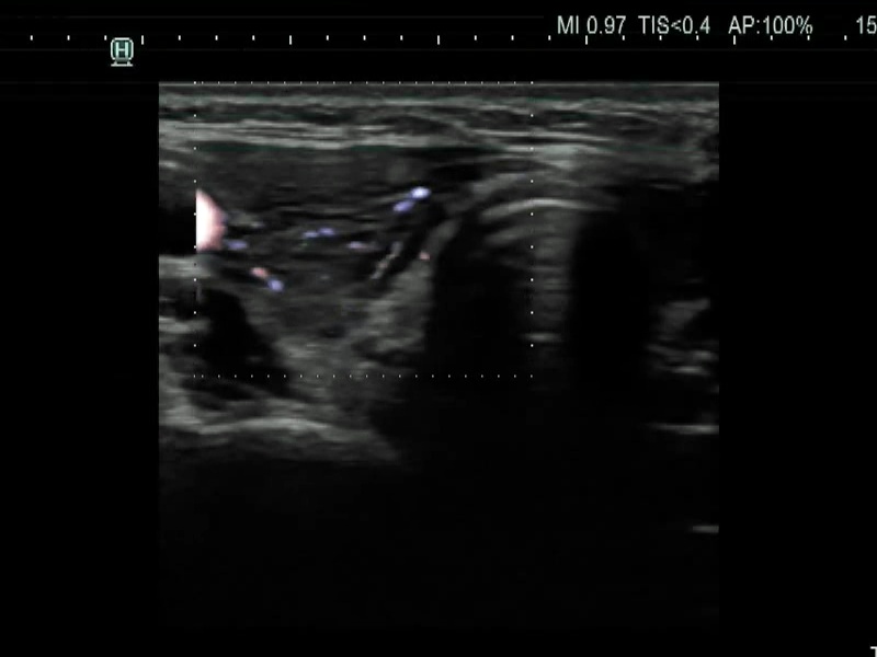
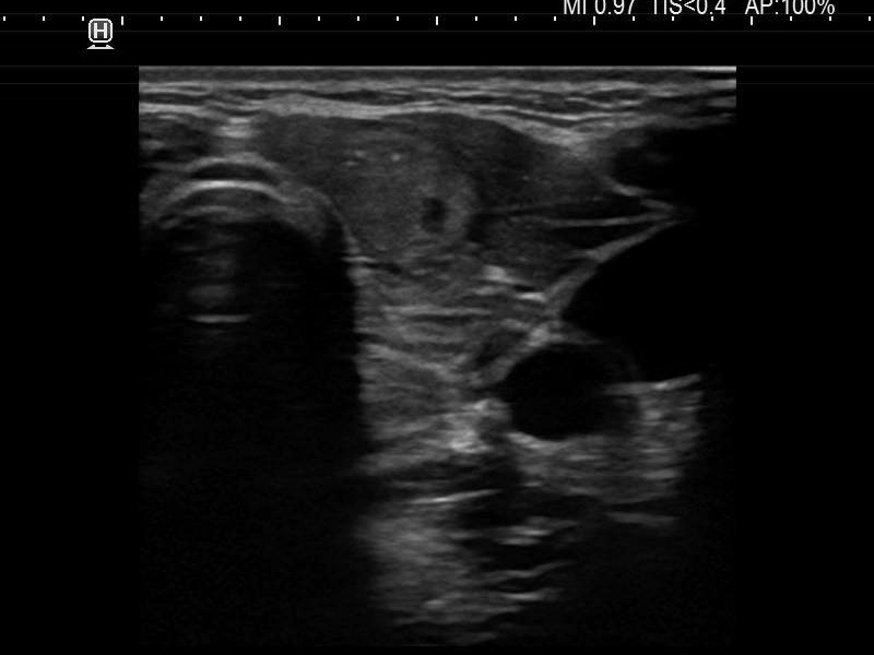
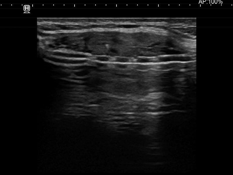
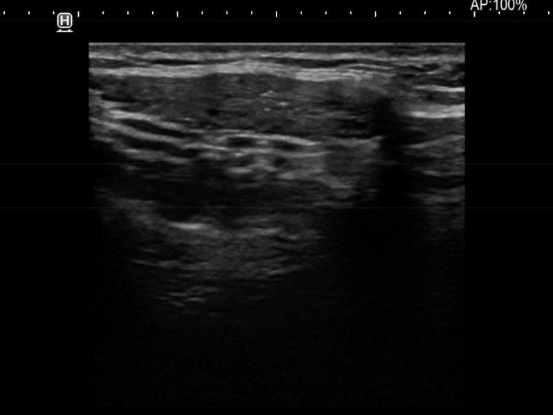
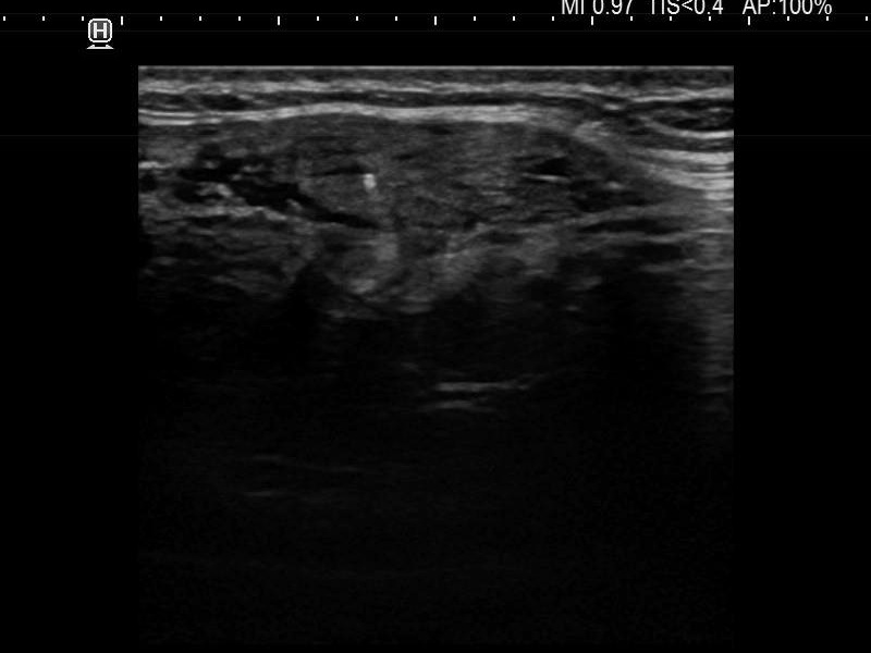
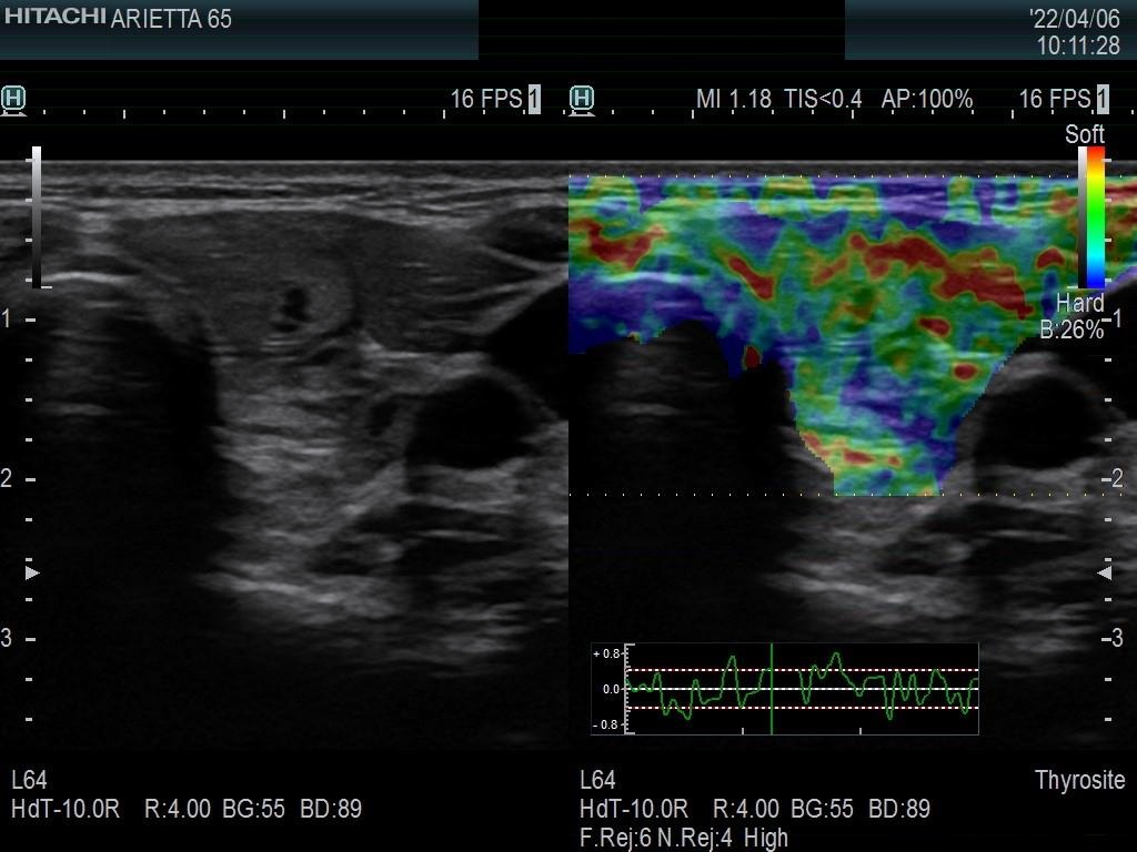
Ultrasonography. The thyroid was moderately hypoechoic and presented with numerous more and less hypoechoic discrete lesions. The largest of the latter was in the lower ventromedial part of the left lobe and had cystic areas and intranodular echogenic figures. The latter included typical comet tail artifacts, back wall cystic figures and some ambiguous small bright granules. The lesion showed both perinodular and intranodular vascularity. Compared with the former examination the nodule increased by 22% in volume. This difference is within the intraobserver variation.
Suggestion: to increase the dose of levothyroxine to daily 150 microgram.
Comment. Based on the presence of typical comet-tail artifacts, the questionable punctate echogenic granules are worth considering also colloid crystals.