
Intranodular hyperechogenic figures - case 808
Follow-up investigation 3 years after the first visit (ultrasonographic picture 6)
|
|

|
|
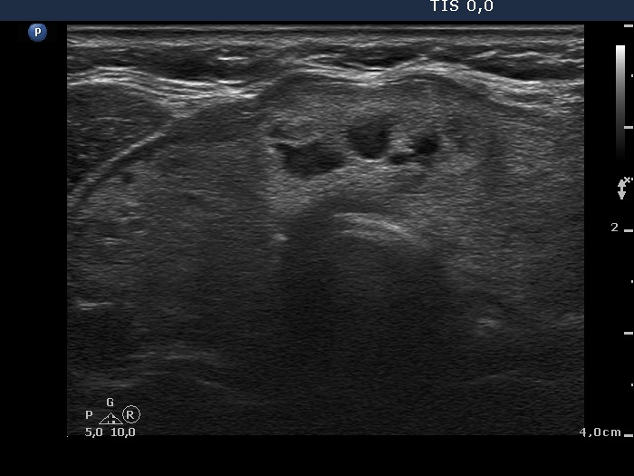
Left side of the isthmus, transverse scan. There is dominantly solid nodule which has some cystic areas.