
Intranodular hyperechogenic figures - case 808 (ultrasonographic picture 3)
|
|

|
|
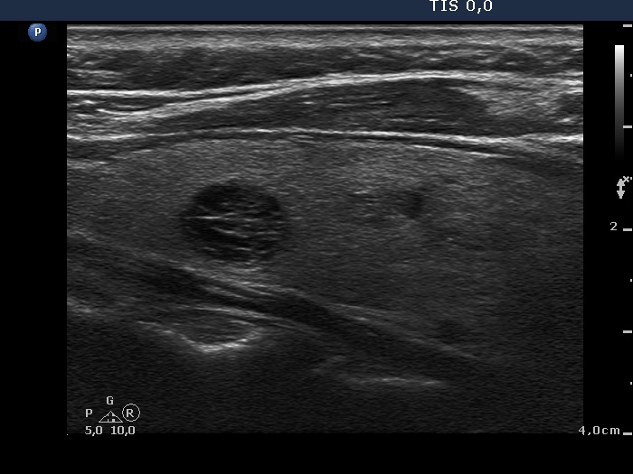
Right lobe, another longitudinal scan. The upper lesion shows a typical presentation of back wall cystic figures: hyperechogenic lines and granules coexist exclusively dorsal to cystic areas.