
Intranodular hyperechogenic figures - case 951
Follow-up examination 2 months later (ultrasonographic picture 5)
|
|

|
|
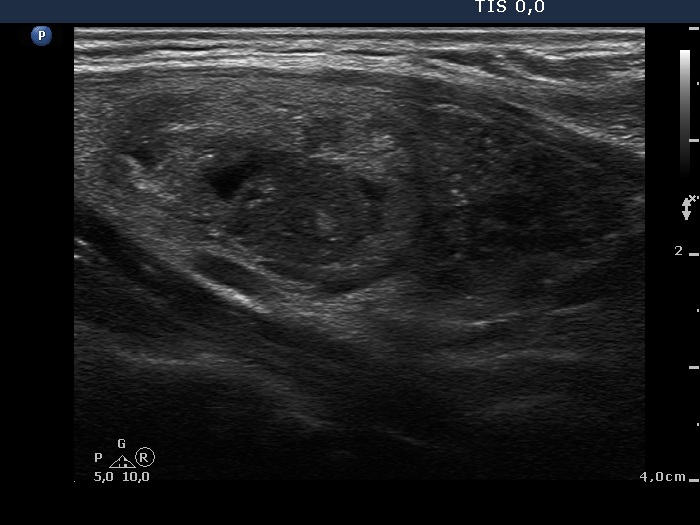
Right lobe, longitudinal scan. Left the upper cystic, right the hypoechogenic nodule.