
Intranodular hyperechogenic figures - case conp 009 (ultrasonographic picture 6)
|
|

|
|
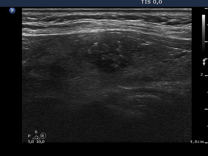
Left lobe, another longitudinal scan. We meet this pattern in every consulting hour: a moderately hypoechogenic lobe presenting discrete hypoechogenic areas - this is the most common ultrasound presentation of Hashimoto's thyroiditis. However, the discrete lesion contains larger and brighter hyperechogenic granules compared with the extralesional part. The figures in the latter are non-specific figures while those within the lesion might be microcalcifications. This difference was the only suspicious sign which suggested that this lesion would be a nodule in a pathological sense and could be even malignant.