
|
|
Elastography - case 1260
|
|

Clinical data: A 79-year-old man was referred for evaluation of nodular goiter detected on preoperative examination of a planned inguinal herniotomy. During the conversation, it was revealed that the patient underwent kidney surgery 14 months ago, one week before the hospital closures in Hungary due to the COVID-19 infection. The patient was not informed of the results of the histological examination. He noticed a lump in the left side of the neck several moths before the present examination.
Palpation: a very hard, not freely moveable mass on the left thyroid lobe. Multiple firm nodes were palpable in the left side of the neck.
Functional state: hypothyroidism on daily 75 microgram levothyroxine with TSH 11.9 mIU/L.
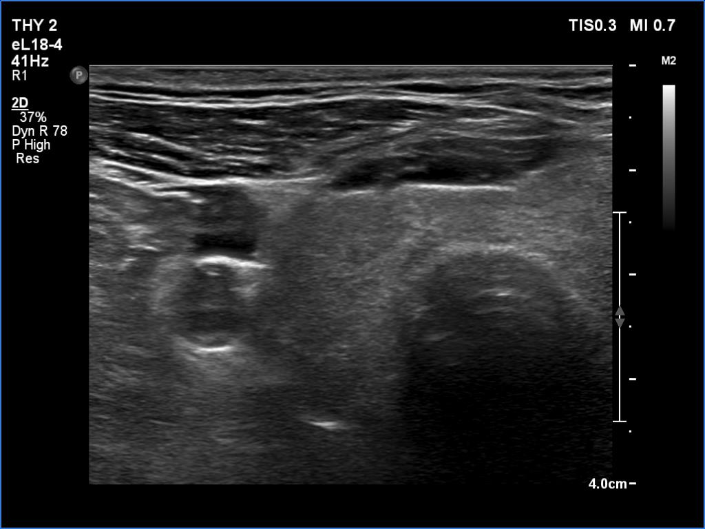
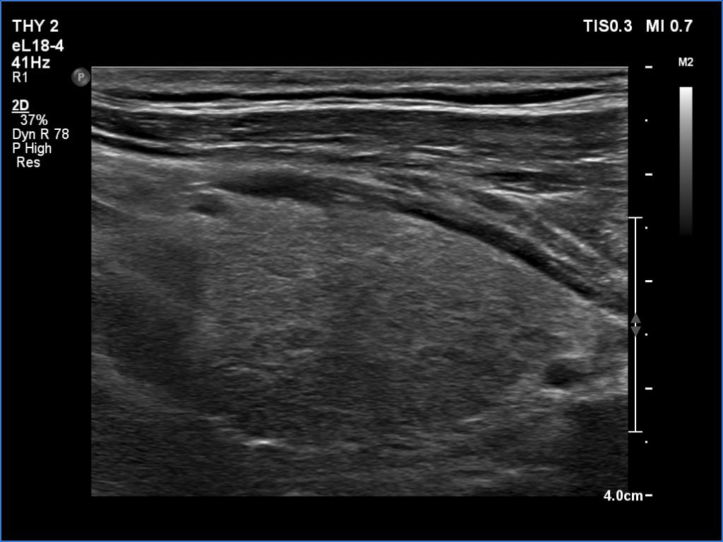
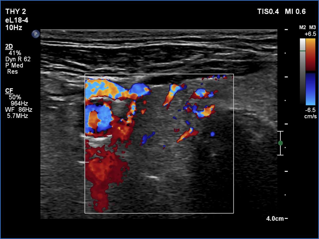
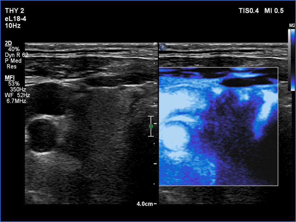
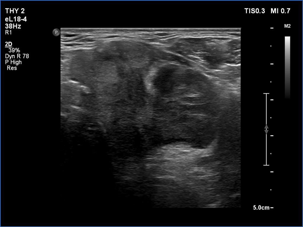
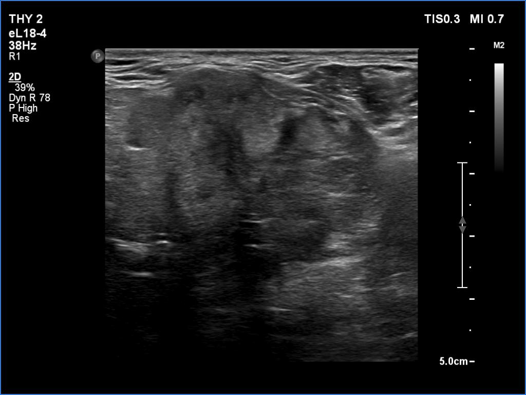
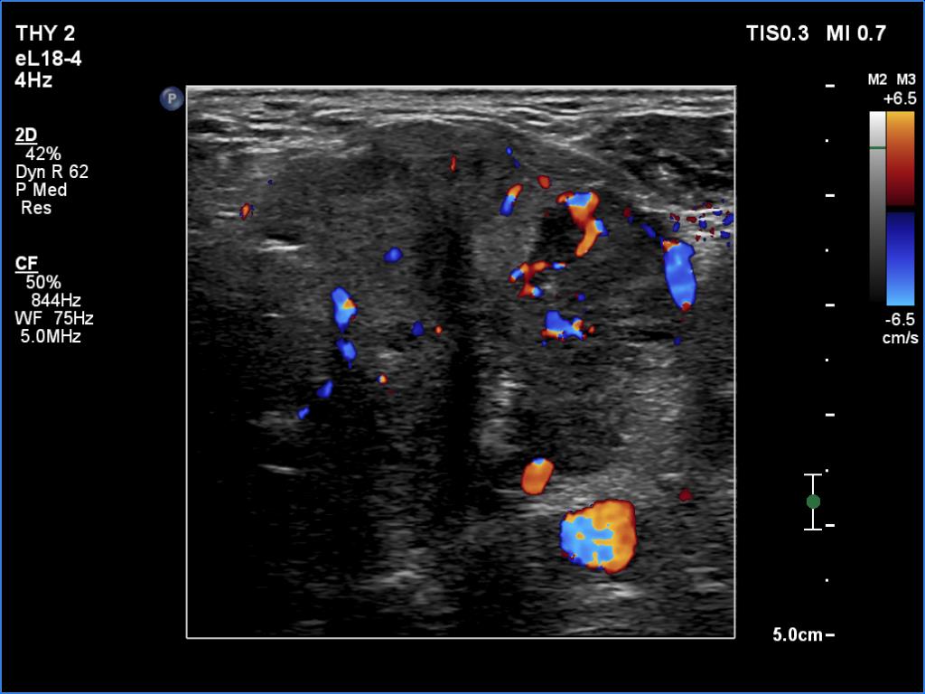
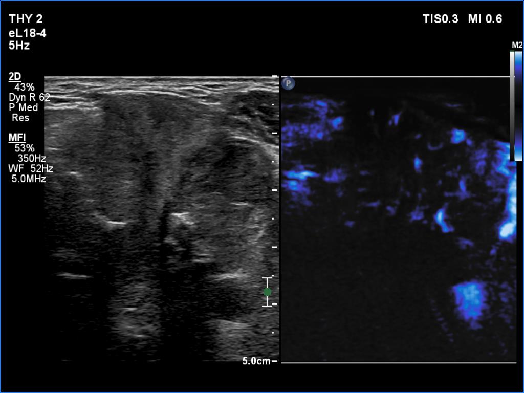
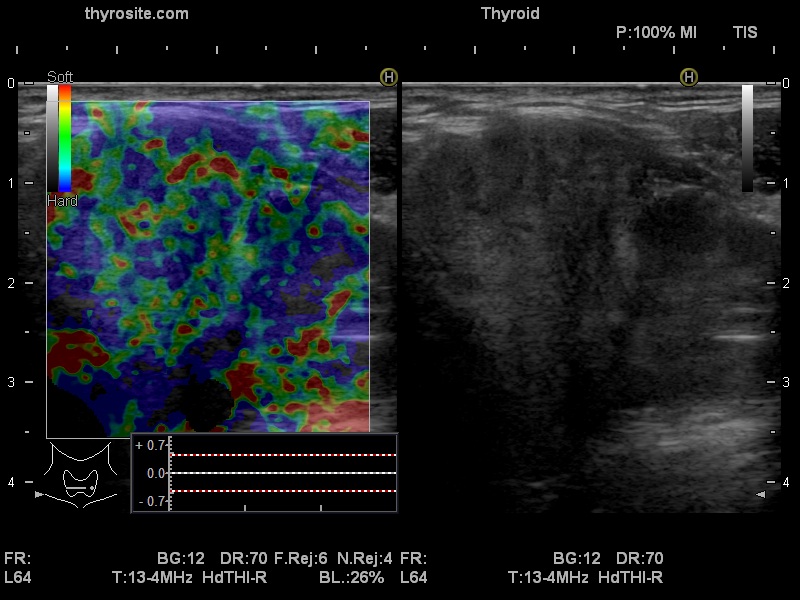
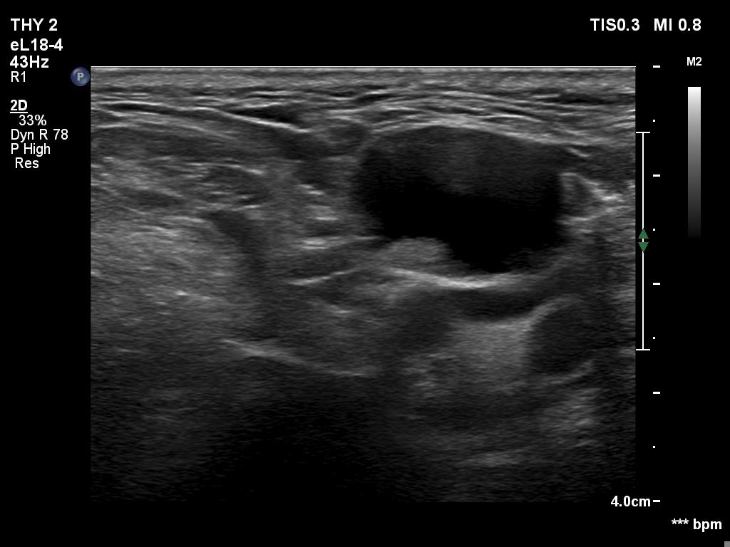
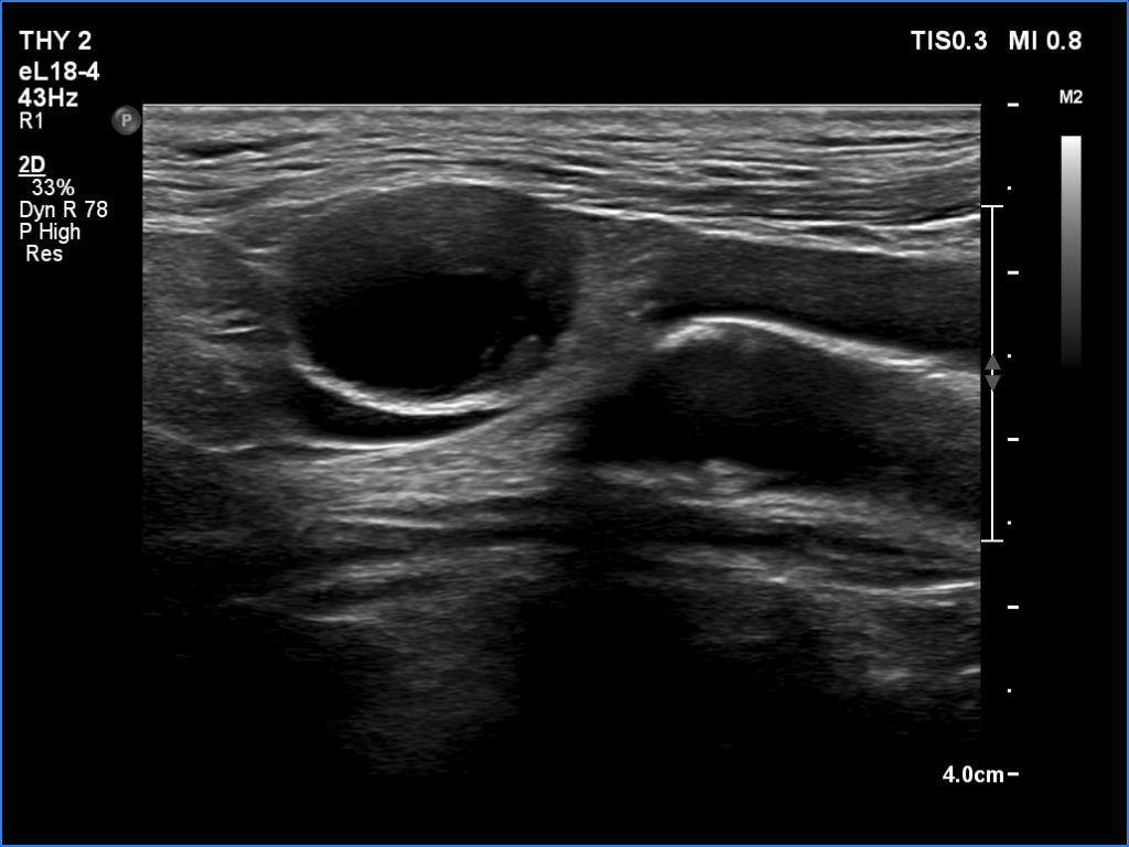
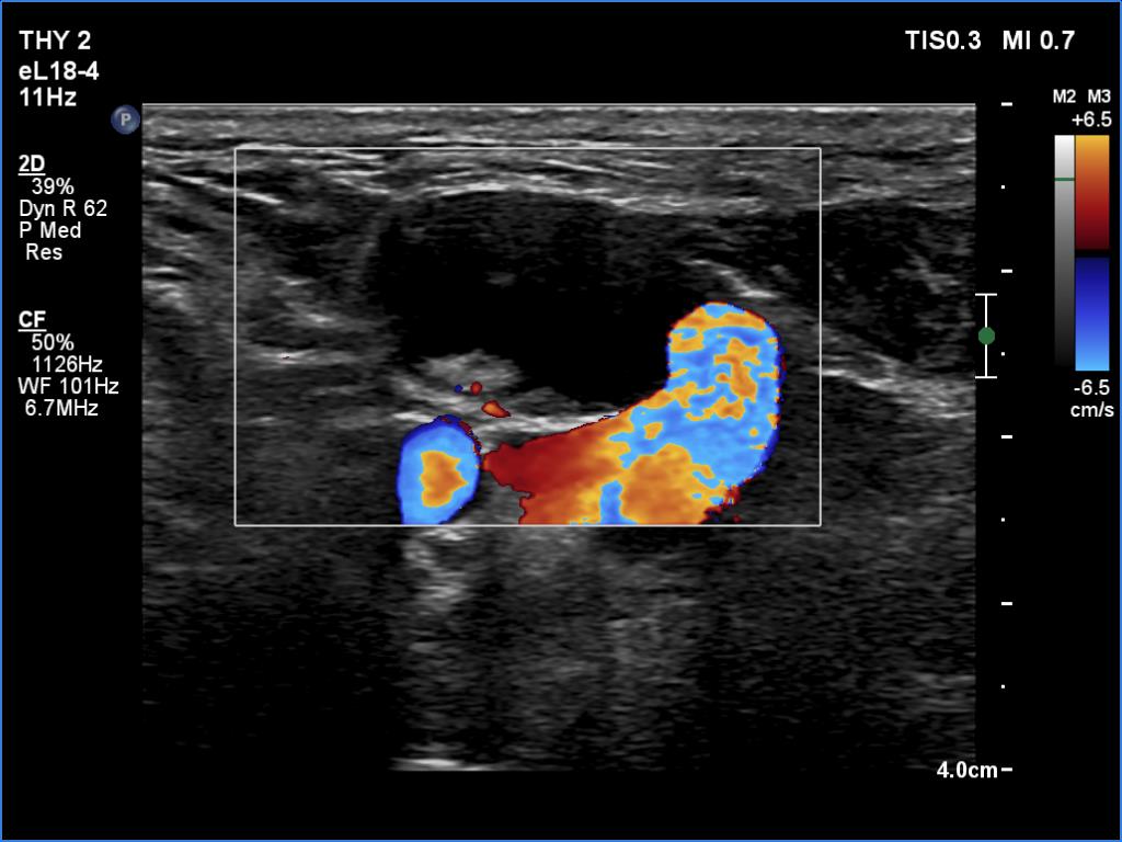
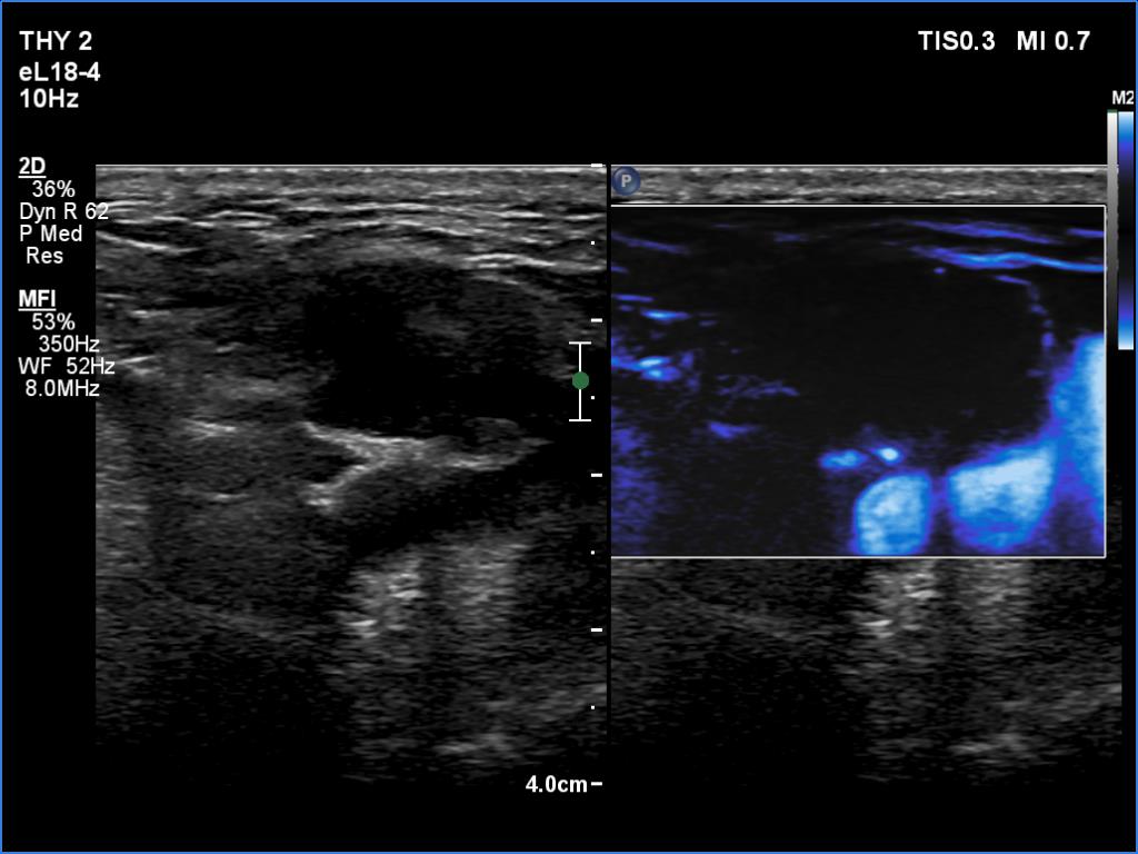
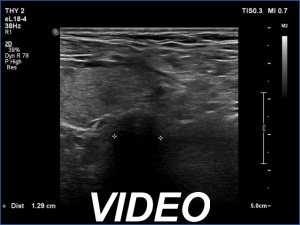
Ultrasonography. The right lobe was echonormal and intact. A large mass occupied almost the entire left lobe. On this side, normal thyroid tissue could only be identified in the dorsal part. The mass was composed of echonormal and hypoechoic areas, had macrocalcifications, irregular intranodular vascularity and proved to be very hard on elastography. There were multiple lymph nodes in III, IV and V left neck compartments. One of the nodes was in close proximity to the jugular vein and was suspicious of having broken into the vessel. The trachea was significantly narrowed at the lower level of the thyroid.
US-guided aspiration was performed from the thyroid mass and from the lymph node presented in the images. Cytology resulted in all three cases in metastatic carcinoma. Wash out thyroglobulin levels were 2.02 and 8.19 ng/L in the thyroid mass and in the neck lymph node, respectively.
After further information, it turned out that the result of the histological examination of the previous surgery was clear cell kidney cancer.
I initiated an oncological council.
Comments.
-
This patient was the victim of the chaos caused by the COVID-19 pandemic.
-
There are several remarkable findings in this case, e.g., the destruction of the left lobe by the tumor, the elastography pattern, the relation of the largest metastatic lymph node and the jugular vein, the ultrasound demonstration of the trachea' stricture.