
Elastography - case 650 (ultrasonographic picture 10)
|
|

|
|
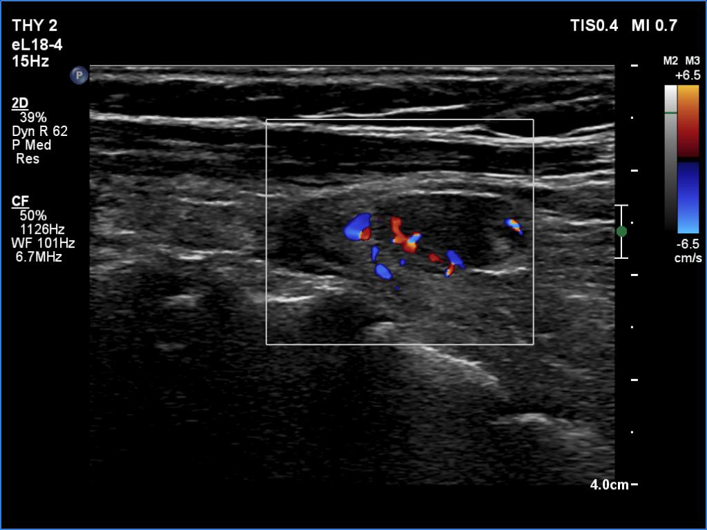
Lower part of the left lobe, longitudinal scan, color Doppler mode. The lesion shows typical presentation of parathyroid vascularity: a vessel antering at apical part and is branching.