
Extrathyroidal spread - case conp 035
Three years prior to present examination (ultrasonographic picture 4)
|
|

|
|
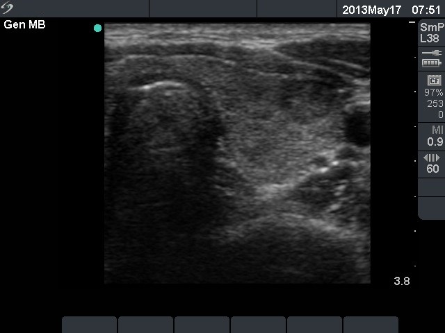
Left lobe, transverse scan. The lobe presents hypoechogenic areas and a larger nodule-like lesion in the ventrolateral part of the lobe. The latter presents microcalcification.