
Extrathyroidal spread - case conp 045 (ultrasonographic picture 5)
|
|

|
|
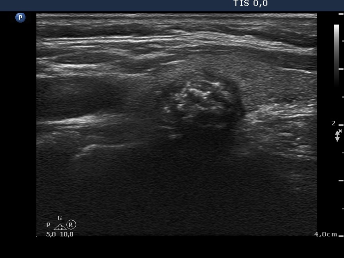
Left lobe, longitudinal view. Three different hyperechogenic figures can be identified within the nodule. The small bright punctate granules are microcalcifications. There are less hyperechogenic punctate and linear figures which correspond to fibrosis. Although we cannot find the primary focus of macrocalcification but the acoustic shadow is the proof for the presence of this structure.