
The shape of the nodule - case 2016 (ultrasonographic picture 6b)
|
|

|
|
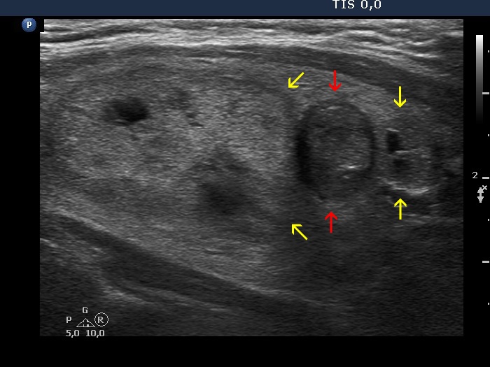
Lower part of the left lobe, longitudinal scan. The hypoechoic nodule (marked with red) is sandwiched between two other nodules (yellow arrows).