
TIRADS - case 1649 (ultrasonographic picture 6)
|
|

|
|
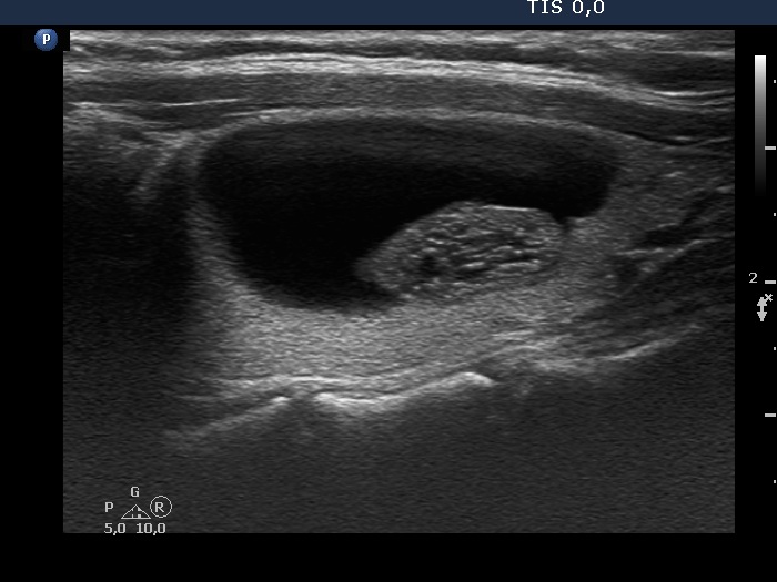
Left lobe, longitudinal scan. The angle between the solid part and the wall of the cyst is acute. Note the presence of hyperechogenic granules within the solid part; all of them are related to a ventral cystic area. Therefore, these figures are caused by posterior back wall cystic enhancement.