
|
|
TIRADS - case 2080
|
|


Clinical presentation: A 39-yr-old woman was referred for evaluation of a nodule described as highly suspicious because of the presence of irregular, partly lobulated, partly spiculated margins and that of microcalcifications. The patient requested the investigation because of complaints suggesting hypothyroidism.
Palpation: Both lobes were firm, but no nodule was palpable.
Laboratory tests: TSH 17.5 mIU/L, FT4 6.5 pM/L, aTPO 36 U/mL.
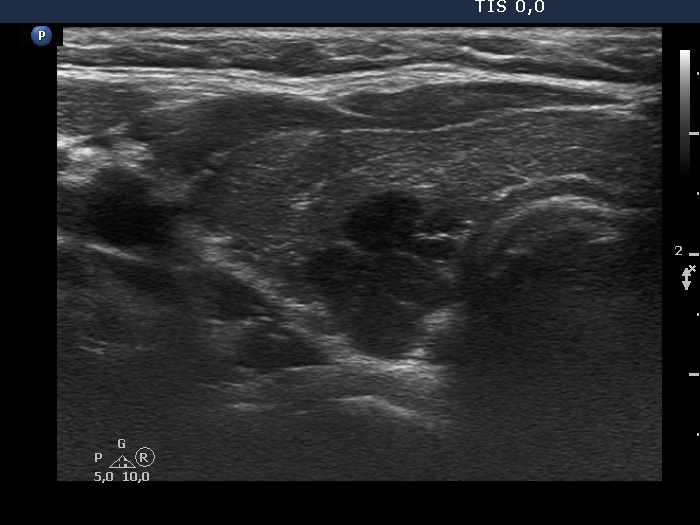
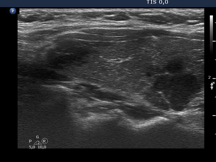
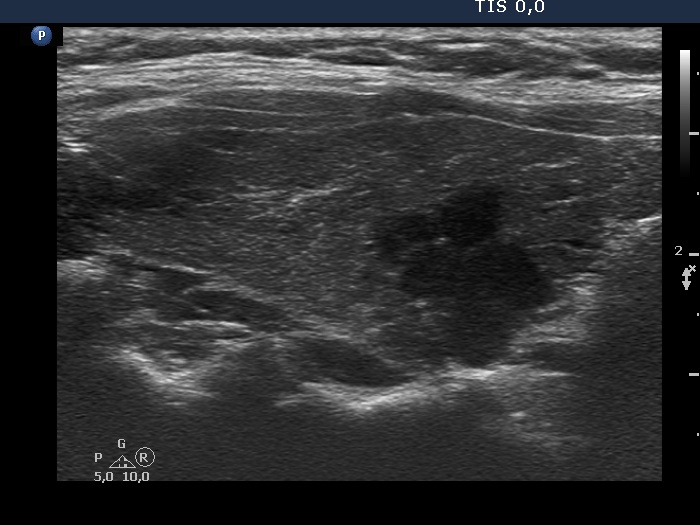
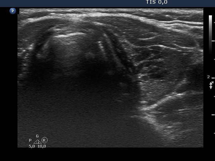
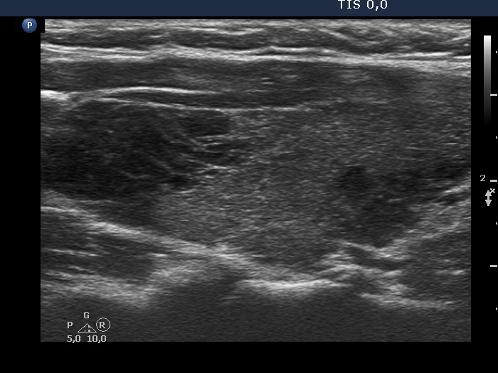
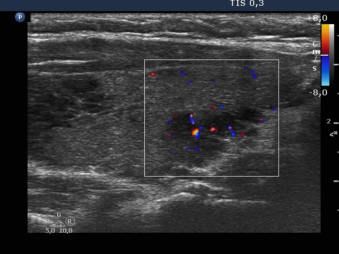
Ultrasonography. The thyroid was minimally hypoechogenic and had several discrete more hypoechogenic areas, including a cluster composed of multiple discrete lesions in the dorsal part of the left lobe. These lesions had irregular, partly lobulated, partly spiculated margins. However, the entire pattern corresponded to Hashimoto's thyroiditis.
Cytology was performed from the lesion located in the dorsal part of the right lobe and resulted in Hashimoto's thyroiditis.
Suggestion: daily 75 microgram levothyroxine therapy.
We met the patient on three more occasions. Despite thorough and repeated explanation of the situation, we failed to fully convince the patient that she is not in danger. Even two years after the initial examination she was afraid harboring thyroid carcinoma. We considered surgical removal of the right lobe.
Histopathology disclosed Hashimoto's thyroiditis without any nodules.
Comment.
-
Regarding the nodule borders the lesions presented partly lobulated, partly spiculated margins. However, these should not be held as pathological nodules, the discrete lesions are presentations of more active foci of Hashimoto's thyroiditis, which is an infiltrative process and therefore frequently has infiltrative, irregular margins.
-
The stigmatizing effect both of an unsubstantiated ultrasound report and that of a high TIRADS score cause great, and depending on the psyche of the patient, even difficult-to-repair harm.
-
To classifying the hypoechoic mass in the right lobe as an EU-TIRADS 5 lesion is not a failure. In this case, it is worth reassuring the patient because the ultrasound pattern stands for thyroiditis. If, on the other hand, the lesion is considered to be EU-TIRADS 1, also in an unquestionable way, it is worth indicating that a cytological examination should be considered despite the classification.
-
The discrete lesions in the left lobe are clearly EU-TIRADS 1 areas.