
|
|
TIRADS - case 294
|
|


Clinical presentation: A 74-yr-old woman was referred for a follow-up examination. We last investigated her five years ago while the first occasion we met her was 23 years ago. At this first visit, a hyperechoic nodule was detected with the dimensions of 11x10x14 mm. Later, two times were aspirated 5 and 9.5 mL cystic content, from the nodule with a maximal diameter of 19 and 26 mm, respectively. Cytology was repeatedly benign.
Palpation: a not firm nodule was suspected in the left lobe.
Laboratory tests: TSH 3.63 mIU/L, aTPO 2 U/mL.
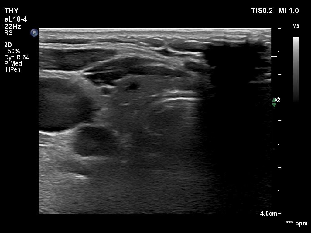
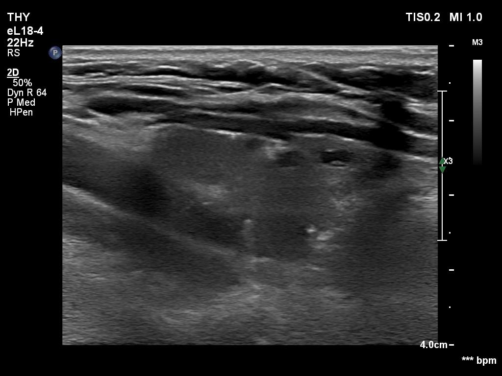
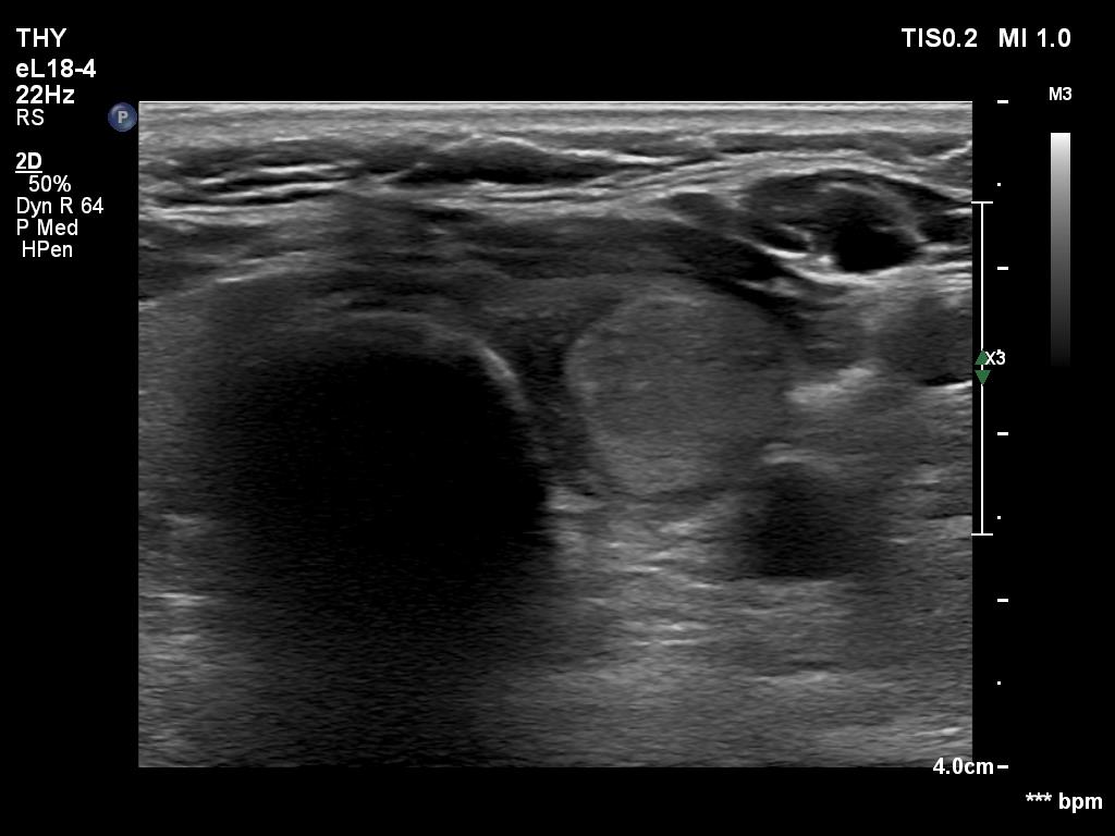
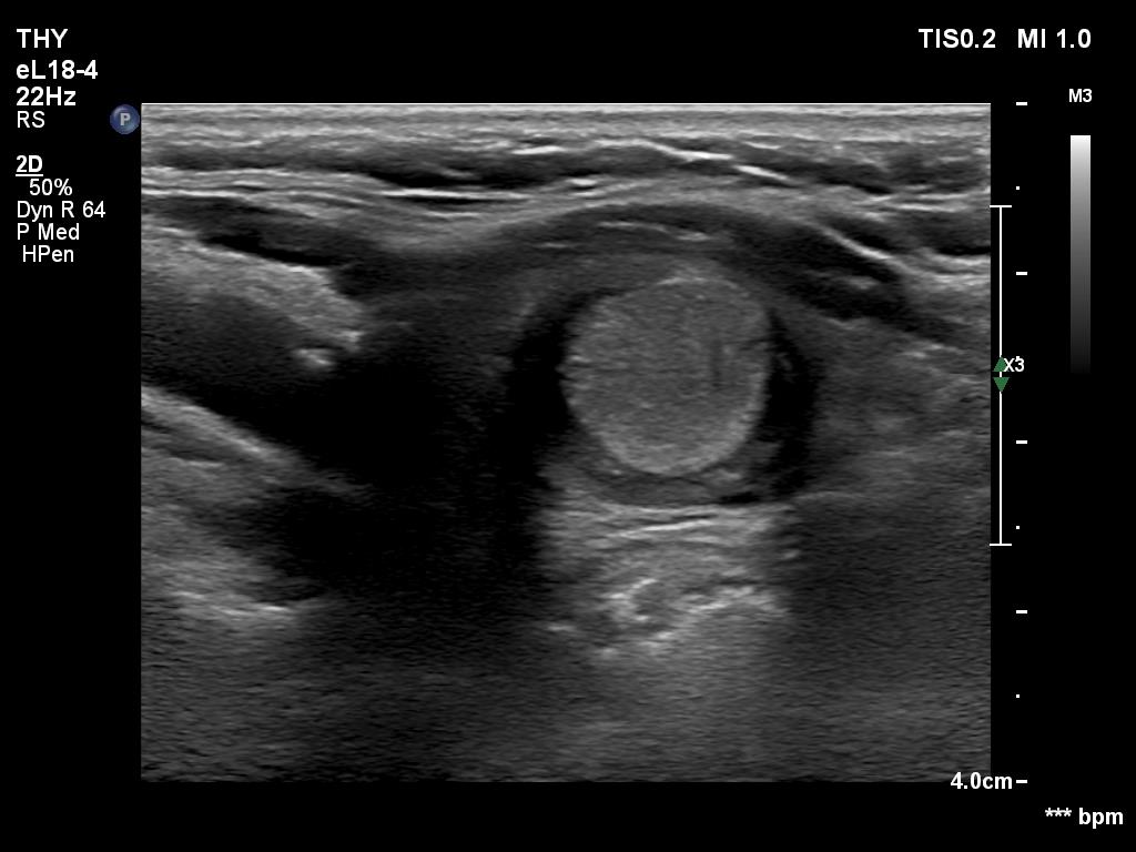
Ultrasonography. The thyroid was minimally hypoechoic. The right lobe has several cystic areas, none of them corresponded to true nodules, these were dilated macrofollicles. There was a dominantly solid nodule in the right lobe. If we compared the echogenicity to a healthy thyroid, then the nodule should be regarded as echonormal, while if the reference tissue was the extranodular part of the lobe, then the nodule should be classified as hyperechoic. The nodule had bright echogenic granule. After a thorough analysis, the most likely explanation was that this was either a back wall figure or comet tail artifact. Nevertheless, microcalcification could not be fully excluded.
Suggestion. Ultrasound in 5 years.
Comment.
-
The discrete cystic lesions in the right lobe are not true nodules, therefore these should be classified as EU TIRADS 1 lesions.
-
The classification of the nodule in the left lobe depends on the interpretation of the bright hyperechoic granule. In my opinion, a single, equivocal bright granule should not be regarded as microcalcification when classifying a nodule. Therefore, I would categorize this nodule as an EU-TIRADS 3 lesion.