
Subacute granulomatous thyroiditis - case 1514
Second examination 3 months later (ultrasonographic picture 5)
|
|

|
|
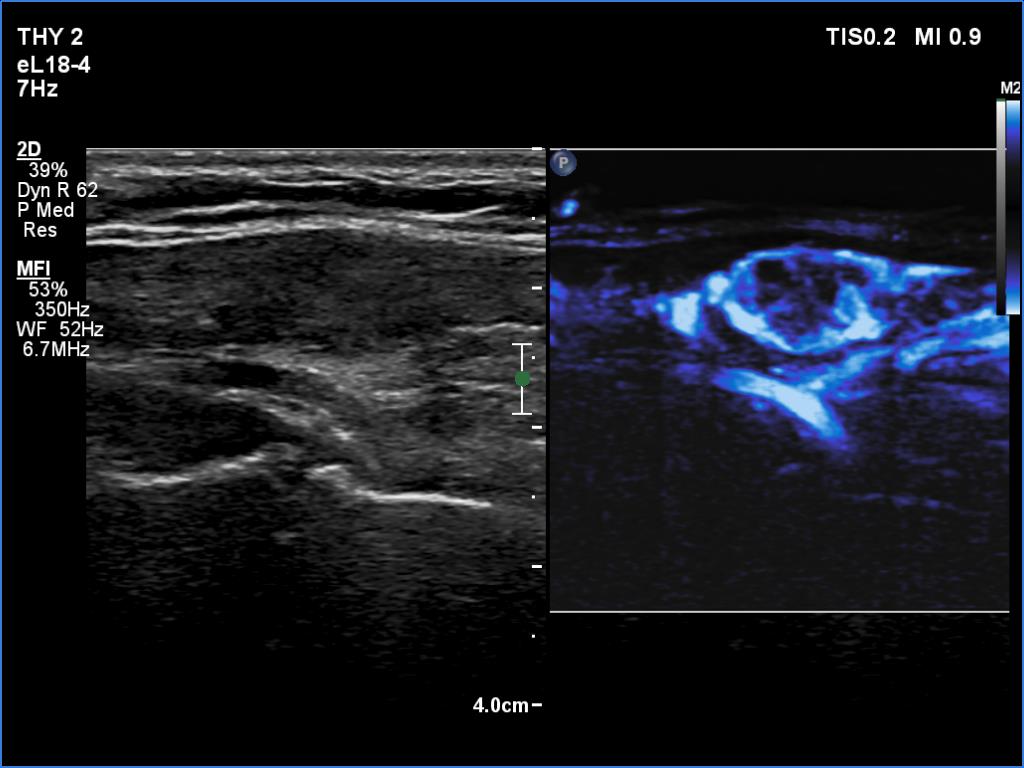
Right lobe, longitudinal scan, microflow imaging. The lesion is rich in vessels while the extralesional part has a decreased vascularity.