
Subacute granulomatous thyroiditis - case 1514
Examination a year after the first visit (ultrasonographic picture 3)
|
|

|
|
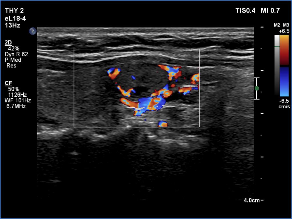
Right lobe, longitudinal scan, color Doppler method. The lesion has perinodular vascularity while the vascularization of the extranodular part is scanty.