
Subacute granulomatous thyroiditis - case 855
Follow-up - 50 months after initial investigation (ultrasonographic picture 6)
|
|

|
|
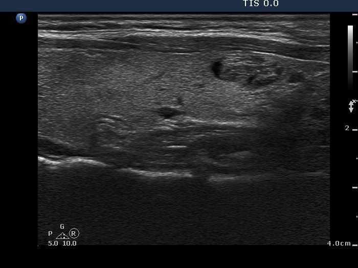
Left lobe, longitudinal scan. In this section, there is a minimally hypoechoic area in the ventral part (left in the image) which corresponds to the previous lesion in question.