
|
|
Lymphocytic thyroiditis - case 1580
|
|

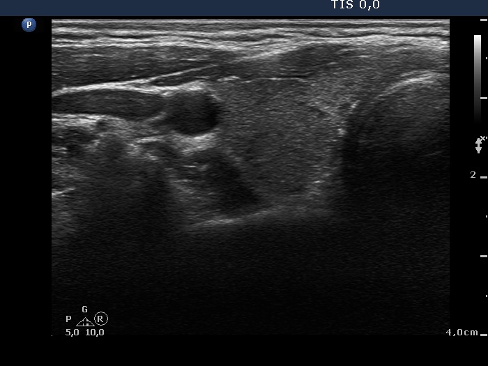
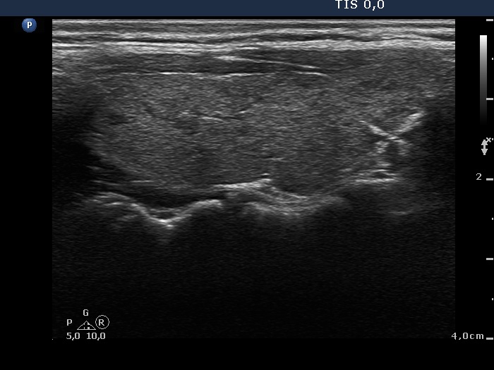
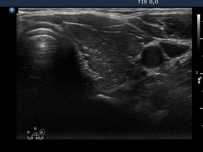
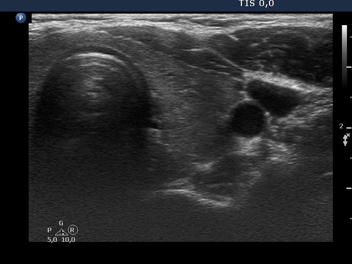
Right lobe, transverse scan

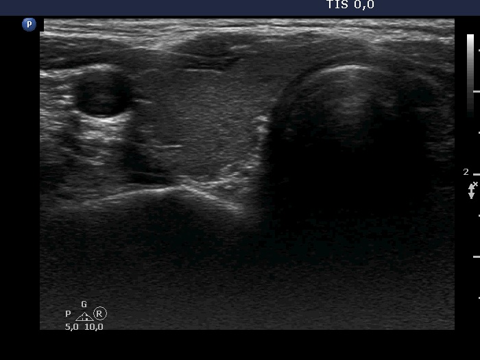
Right lobe, longitudinal scan


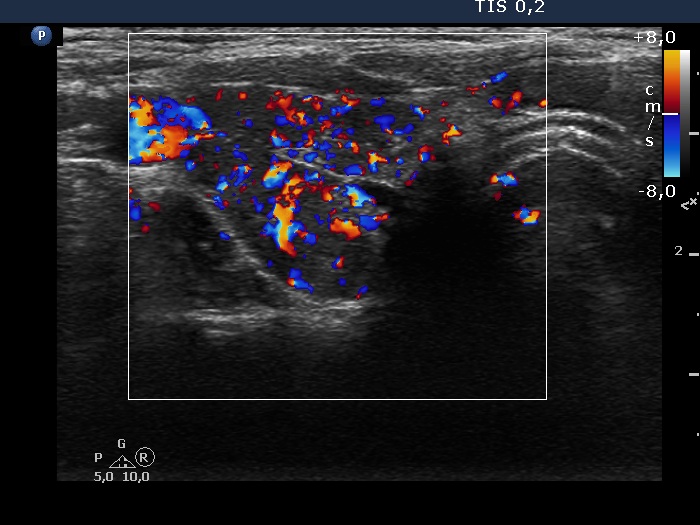
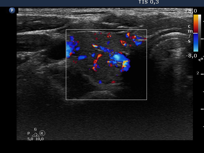
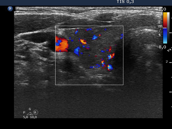
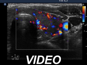
Right lobe, color Doppler mode

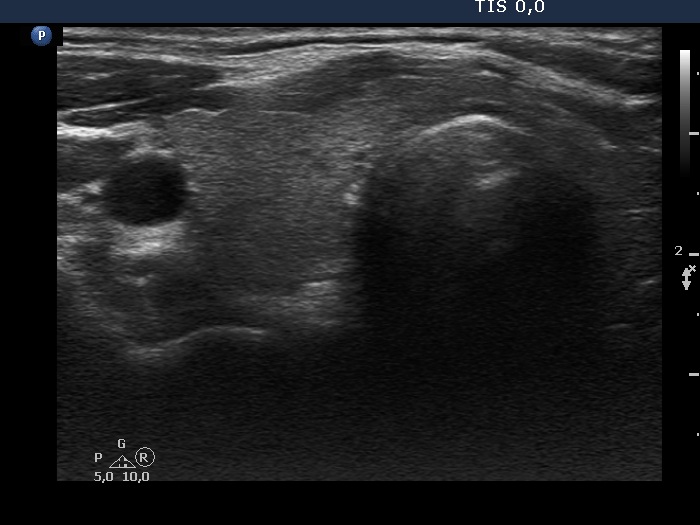
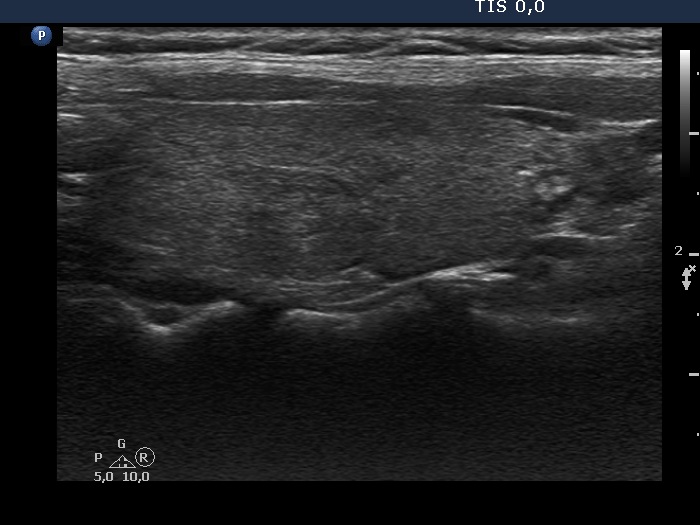
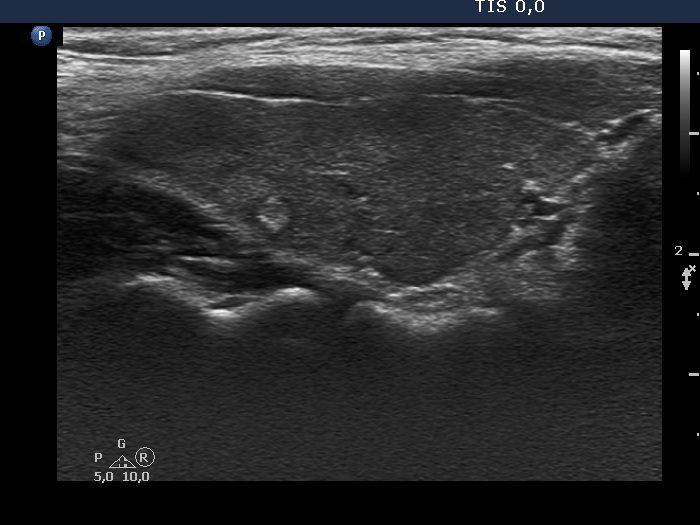
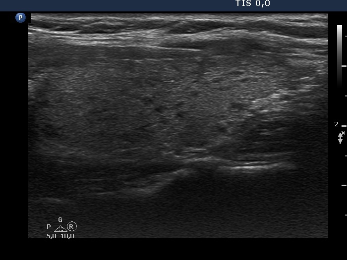
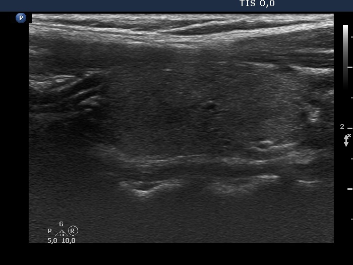
Left lobe, transverse scan


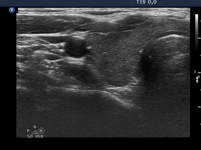
Left lobe, longitudinal scan


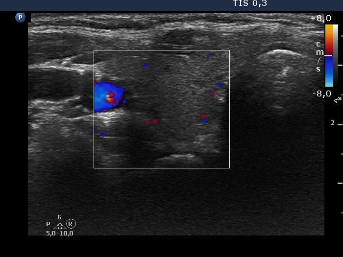
Left lobe, color Doppler mode


Clinical data: A 33-year-old woman requested an evaluation of a thyroid dysfunction. The patient gave birth 5 months ago. Two months before the present study, the TSH was < 0.001 mIU/L, the FT4 was 26.9 pM/L. She has not taken any medicine so far for thyroid disease. She experienced fatigue, palpitations and hair loss.
Palpation: no abnormality.
Laboratory tests: TSH < 0.001 mIU/L, FT4 15.5 pM/L, aTPO 302 U/mL, TSAb < 0.3 U/L.
Ultrasonography. The thyroid was echonormal and presented moderately hypoechoic areas. The echogenicity index was around 15%. The vascularity was decreased.
Suggestion: beta-blocking drug as required. Repeat hormonal evaluation in 3 months.
Summary of ultrasound and laboratory data:
Date of examination |
Volume of the thyroid (mL) |
Basic echogenicity |
Vascularity |
Results of laboratory investigations |
|
| Right lobe |
TSH (mIU/L) |
FT4 (pM/L) |
|||
5 months after delivery |
8.8 |
Echonormal |
Low |
< 0.001 |
15.5 |
9 months after delivery |
19.1 |
Deeply hypoechoic |
Increased |
18.0 |
7.9 |
12 months after delivery |
10.8 |
Moderately hypoechoic |
Average |
3.0 |
9.3 |
4 months after delivery |
9.9 |
Minimally hypoechoic/echonormal |
Very low |
0.01 |
14.7 |
11 months after delivery |
9.8 |
Minimally hypoechoic |
Average |
3.7 |
9.2 |
Comment.
Post partum thyroiditis could be detected after two deliveries in this patient. It is worth noting that both biochemical and ultrasound data were very similar at the same time after deliveries, i.e. at the first and fourth and at the third and the fifth examination.