
Lymphocytic thyroiditis - case 41
Follow-up investigation 2 years later (cytologic picture 6)
|
|

|

|
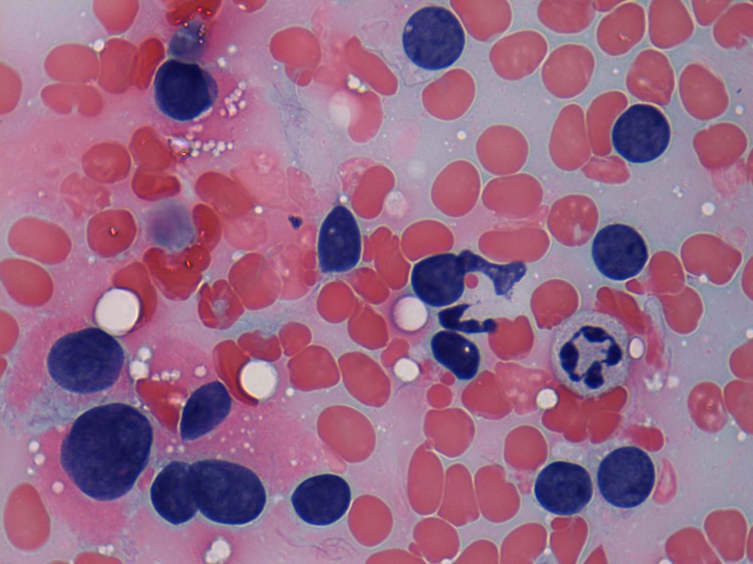
Pap-smear, 1000x. Typical oxyphilic cells are demonstrated in the left part of the image. The cytoplasm of Hürthle-cells is finely granular.