
|
|
Lymphocytic thyroiditis - case 609
|
|

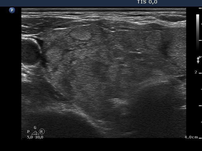
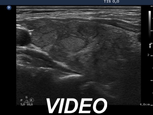
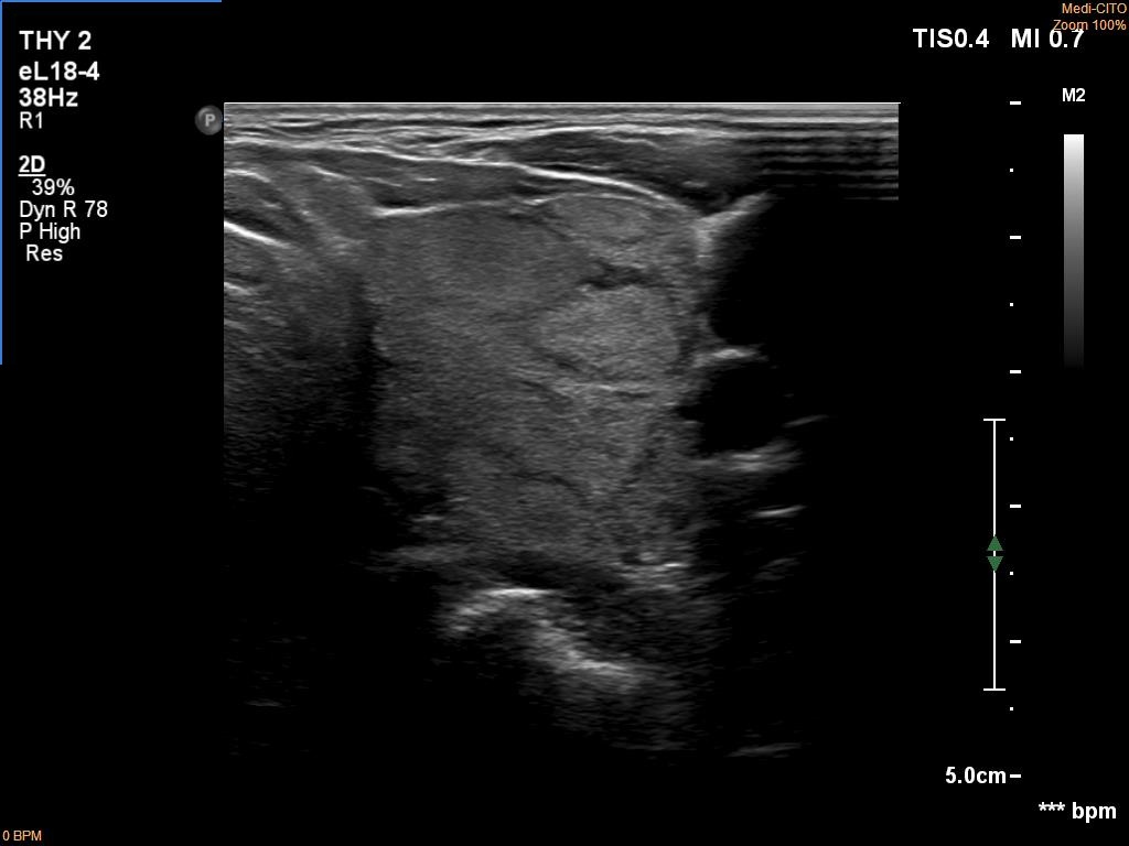
First examination (first row of images):
Clinical data: A 75-year-old woman was referred for evaluation of a goiter which was palpated by her GP.
Palpation: Both lobes were a bit firm and enlarged. There was a firm nodule in the isthmus.
Laboratory tests: TSH 4.49 mIU/L, aTPO 40 U/mL.
Ultrasonography. The thyroid was echonormal and had numerous discrete echonormal lesions. The largest of them was located in the isthmus. None of the lesions shared oncological importance.
Cytology was performed form the lesion in the isthmus and resulted in lymphocytic thyroiditis.
Suggestion: TSH and ultrasound in a year.
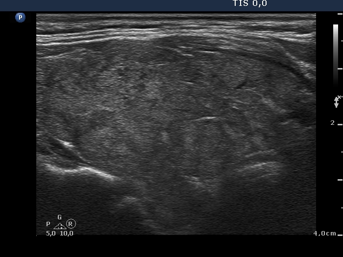
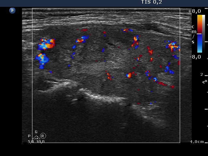
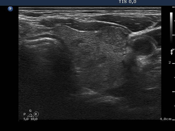
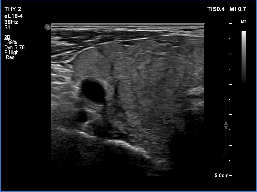
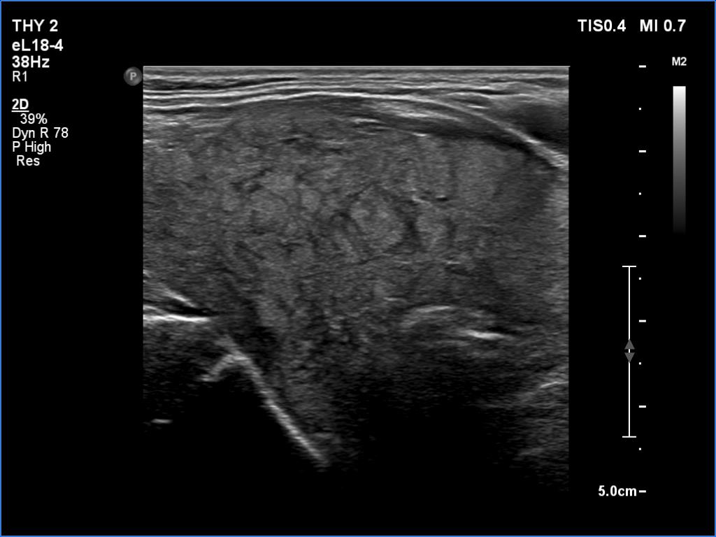
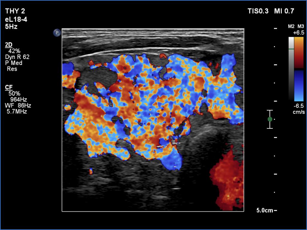
Second examination 6 years later (second row of images):
Clinical data: The patient was referred for a follow-up.
Palpation: unchanged.
Laboratory tests: TSH 3.52 mIU/L.
Ultrasonography. The pattern was the same as in the previous study except for the lesion in the isthmus which has been increased in size.
Suggestion: TSH in a year, ultrasound in 3 years.
Comment. This is the typical presentation of the micronodular form of Hashimoto's thyroiditis.