
Lymph nodes - case 1131 (ultrasonographic picture 13)
|
|

|
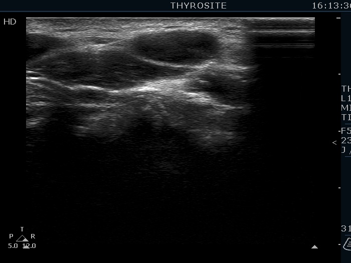
Above and lateral to the left lobe, longitudinal scan. The hypoechoic mass lower and ventral to the muscle fiber has a pale linear figure which might correspond to hilum.