
|
|
Parathyroid lesions - case 110
|
|

Clinical data: A 38-year-old man was referred for evaluation of hyperparathyroidism. On routine blood test, elevated calcium and decreased phosphate levels were found. Thereafter, parathormone level proved to be increased. The patient had no complaints.
Palpation: no abnormality.
Laboratory tests: TSH 1.08 mIU/L, serum-parathormone 120.7 pg/mL (normal value 12-88), serum calcium 2.81 mM/L, phosphorus 0.73 mM/L.
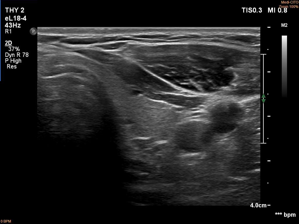
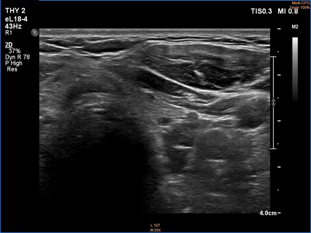
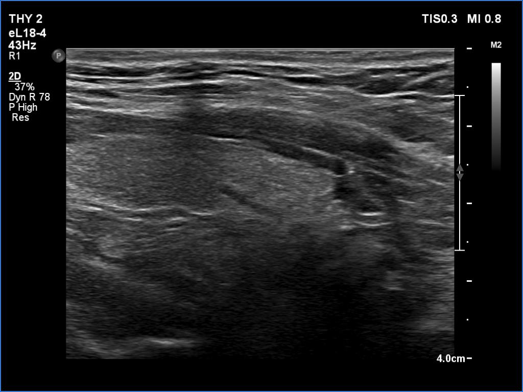
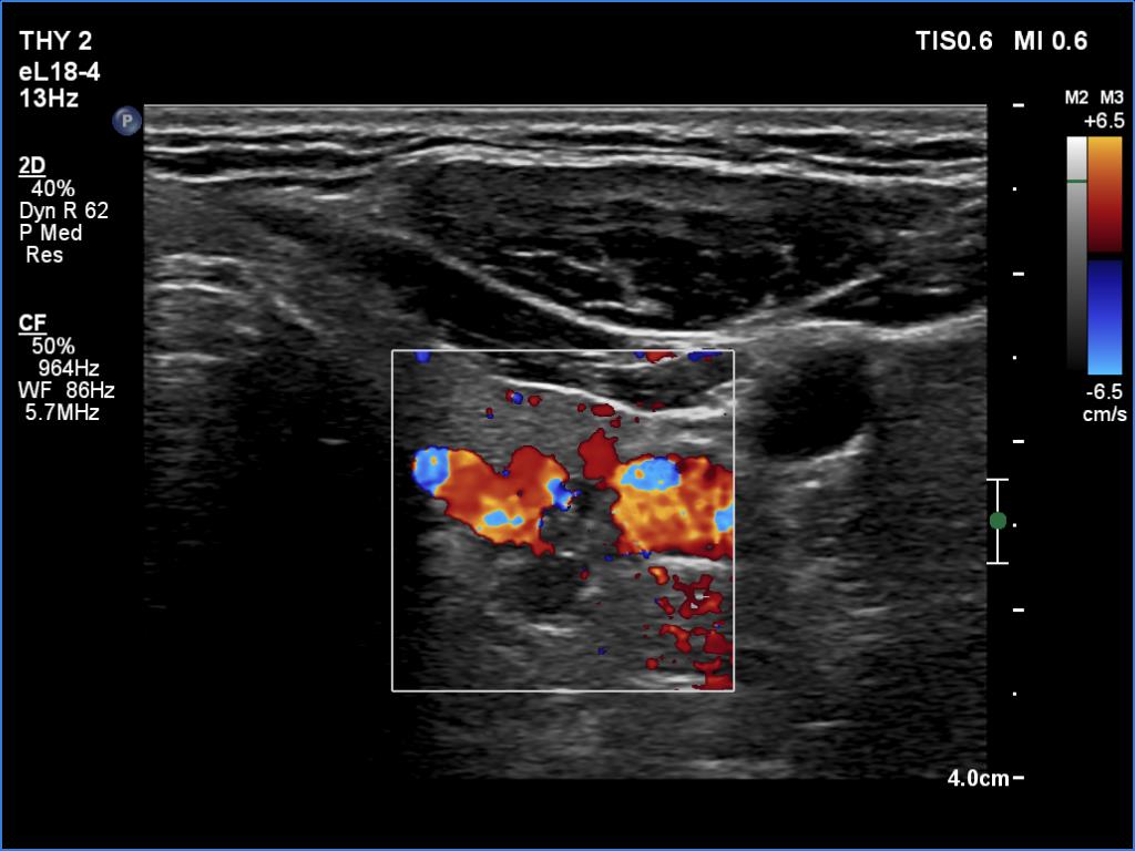
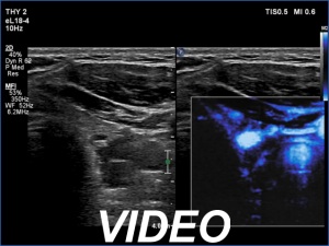
Ultrasonography. The thyroid was echonormal. There was a hypoechoic mass dorsal and under to the lower pole of the left thyroid lobe.
We tried to gain cytological material two-times from the mass but it was doubtful whether we reached the hypoechoic mass. Aspiration cytology was non-diagnostic. Wash-out parathormone was 0.1 pg/ml.
Scintigraphy disclosed parathyroid adenoma corresponding to the left lower parathyroid.
Histopathology: left lower parathyroid adenoma.
Comment. It is more difficult to gain adequate cytological material from a deep lesion than from a more ventral one.