
Parathyroid lesions - case 742 (ultrasonographic picture 4)
|
|

|
|
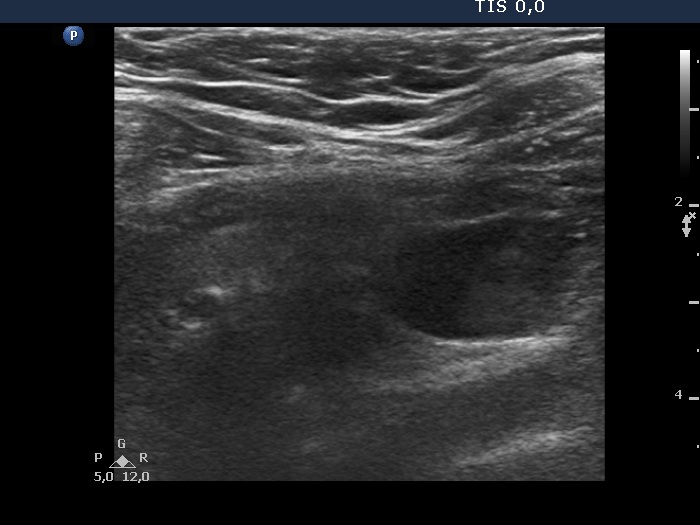
Left lobe, longitudinal view. It was impossible to place the transducer on the patient's neck so that both the thyroid gland (upper, i.e. left in the image) and the parathyroid gland (lower, i.e. right in the image could be well represented.