
Teamwork - case 1091
First examination (ultrasonographic picture 4)
|
|

|
|
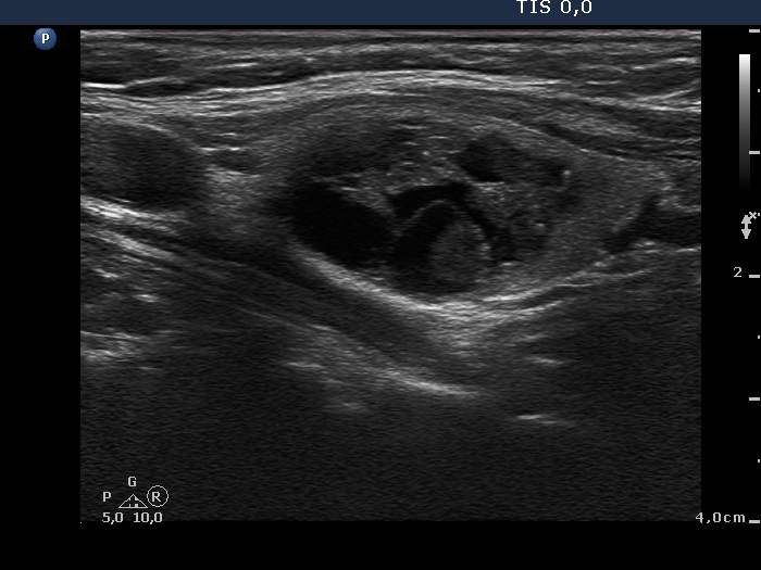
Left lobe, another longitudinal scan. In this section we can reveal intranodular hyperechogenic lines dorsal to cystic areas, therefore these intranodular hyperechogenic figures are caused by back wall posterior enhancement.