
Teamwork - case 201
Two years after the initial examination (ultrasonographic picture 4)
|
|

|
|
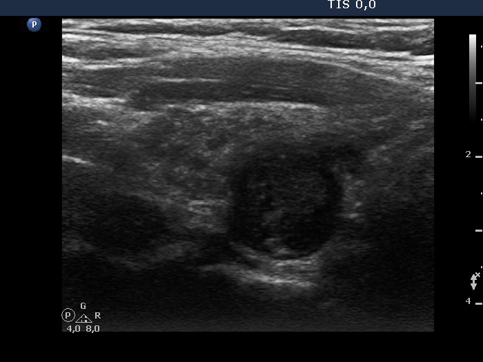
Right lobe longitudinal view - correct settings. Compared with the tiny hypoechoic areas, the dorsal lesion has a more regular shape, and it is more hypoechoic.