
|
|
Teamwork - case 2178
|
|


Clinical presentation: A 42-yr-old woman requested follow-up evaluation of a thyroid cyst which has been known for 7 years.
Palpation: The right lobe was suspicious having a soft nodule.
Laboratory tests: TSH 1.74 mIU/L.
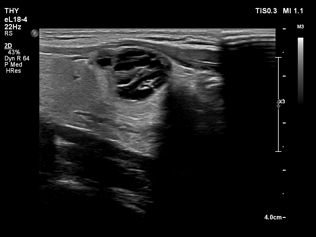
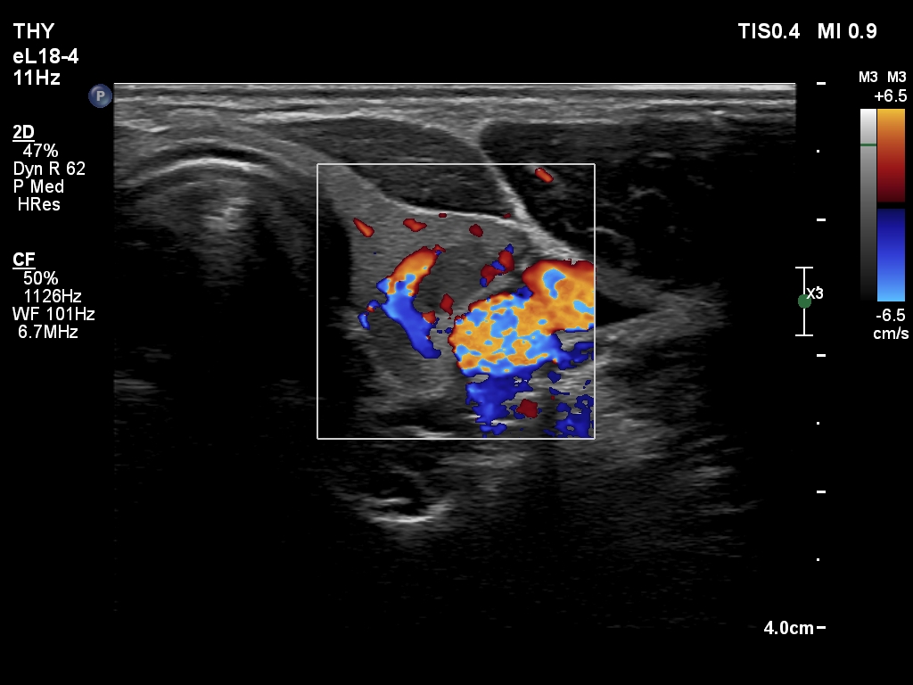
Ultrasonography. The thyroid was echonormal. There was a cystic nodule in the right lobe. The lesion had spongiform portions. The nodules presented all three possible signs of an extrathyroidal extension. There was a minimally hypoechoic nodule in the left lobe.
Cytology of the nodule in the left lobe resulted in benign follicular proliferation.
Suggestion: ultrasound in 5 years.
Comment.
- The categorization of the cystic nodule depends on the definition of spongiform nodules: the spongiform areas exceeded 50% but the nodule was not entirely spongiform.
- Great proportion of benign nodule presents signs of a possible extrathyroidal extension because these features are non-specific, this is particularly true for cystic nodules located at the edge of a lobe. The liquid produced often bulges the surface of the thyroid. The contour of this nodule was abutting and bulging and the thyroid capsule was discontinuous.
- The small nodule presented irregular shape but this was not of pathological importance. The hard wall of the carotid artery prevented the nodule from spreading sideways.