
|
|
Case 1298/only for Videolibrary and Exam/ |


Clinical data: A 66-year-old woman was referred for evaluation of a suspicious nodules which was found on carotid Doppler examination. The patient has been treated for hypothyroidism for more than 10 years.
Palpation: no abnormality.
Laboratory test: TSH 6.02 mIU/L on daily 100 microgram levothyroxine.
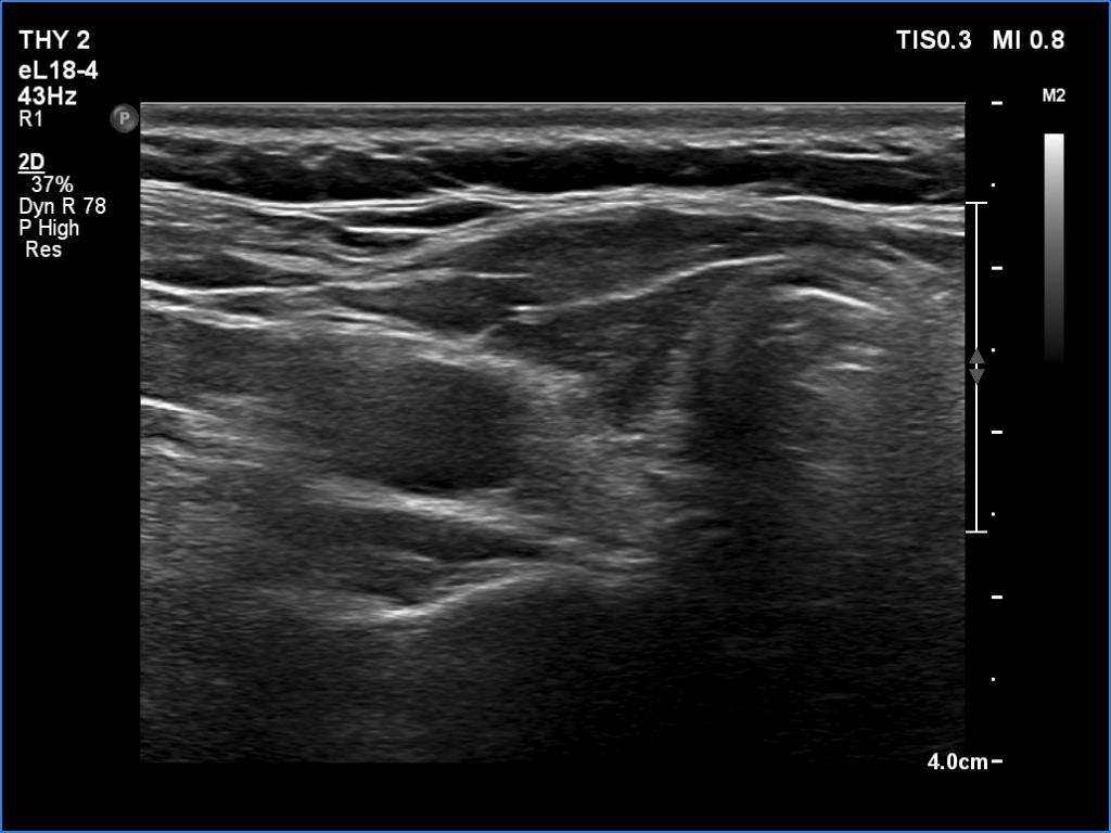
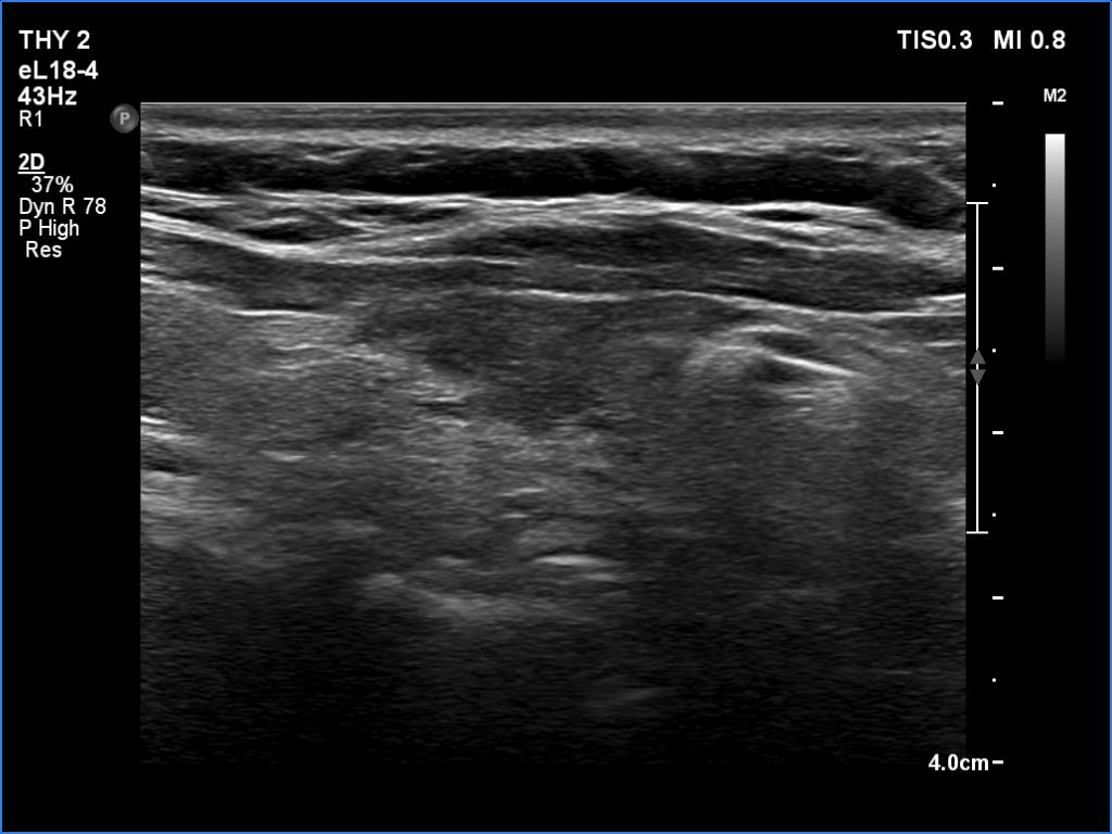
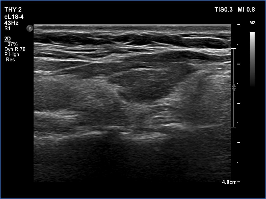
Ultrasonography. The thyroid was atrophic. Both lobes were composed of a central, larger hypoechoic area surrounded with echonormal tissue. The pattern did not correspond to nodule. The vascularity was decreased.
Cytology was performed form the right lobe and resulted in Hashimoto's thyroiditis.
Suggestion: daily 125 microgram levothyroxine.
Comment. A central hypoechoic part surrounded with echonormal tissue is one of the typical presentations of Hashimoto's thyroiditis, not infrequently misinterpreted as a large hypoechoic nodule which occupies great part of a lobe. The irregular margins, the infiltrative borders of the thyroiditis are the main clues of differentiation this presentation from nodular goiter. The pattern can be observed usually in both lobes which is also characteristic of this form of thyroiditis.