
Case 1348
Second examination 3 months later (ultrasonographic picture 2)
|
|

|
|
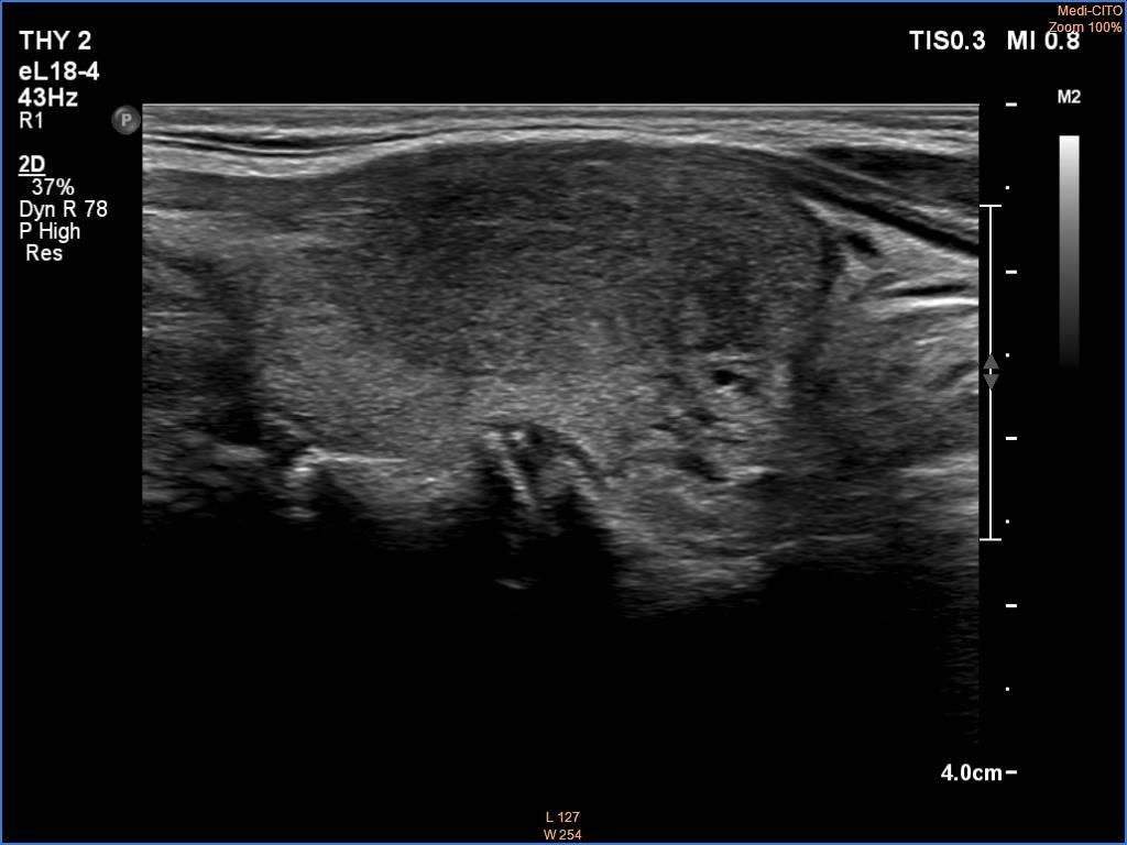
Right lobe, longitudinal scan. As in most case of discrete lesions of thyroiditis, the borders are non-regular but partly blurred, partly lobulated.