
|
|
Case 1441/only for Videolibrary and Exam/ |
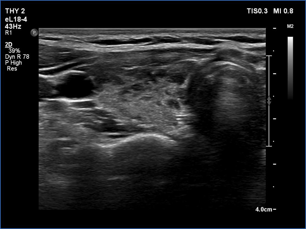
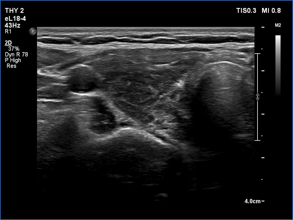
First examination (first row of images):
Clinical data: A 26-year-old woman at the 8th gestational week was referred for evaluation. She has been treated for hypothyroidism for 5 years.
Palpation: Both lobes were firm on palpation. No nodule could be palpated.
Laboratory tests: TSH 6.24 mIU/L, FT4 10.1 pM/L on daily 50 microgram levothyroxine.
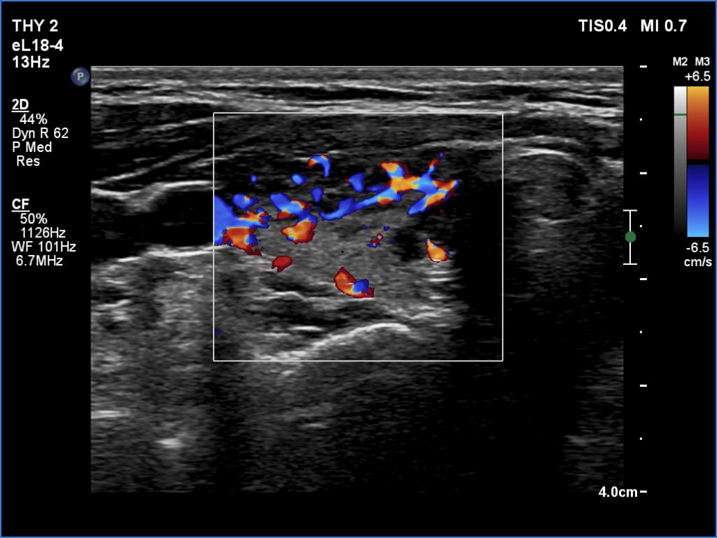
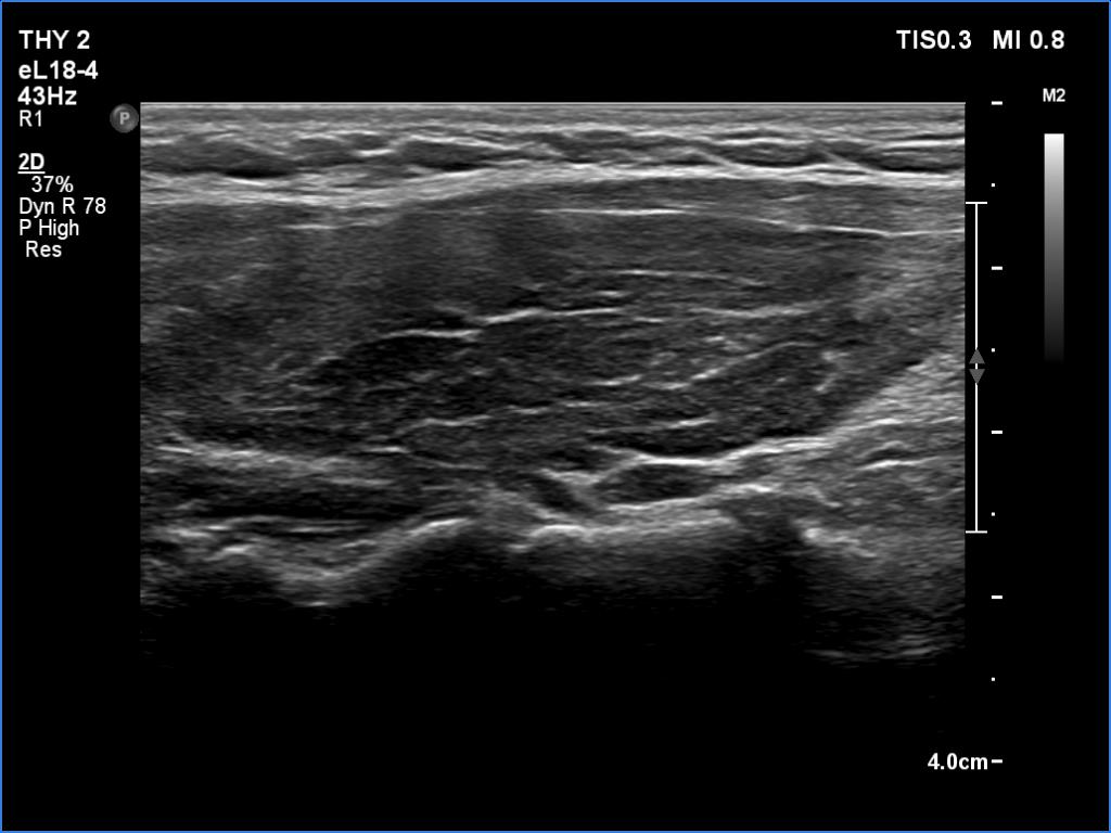
Ultrasonography. The thyroid was echonormal and presented numerous hypoechogenic discrete areas. The echogenicity index was around 30% and 60%, right and left lobe, respectively. None of the discrete areas corresponded to pathological nodule.
Suggestion: to increase the dose of levothyroxine to daily 75 micrograms. Repeat TSH in 6 weeks.
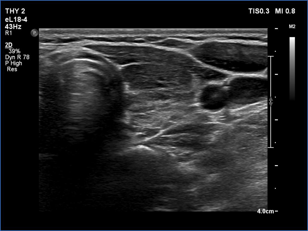
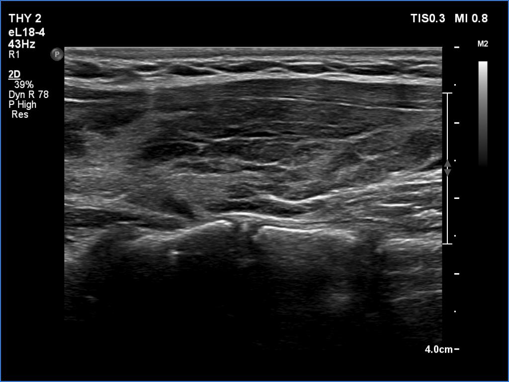
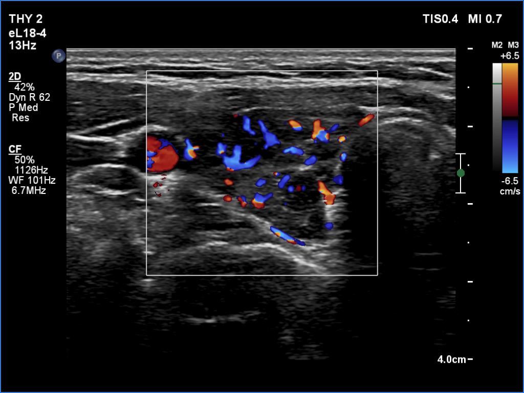
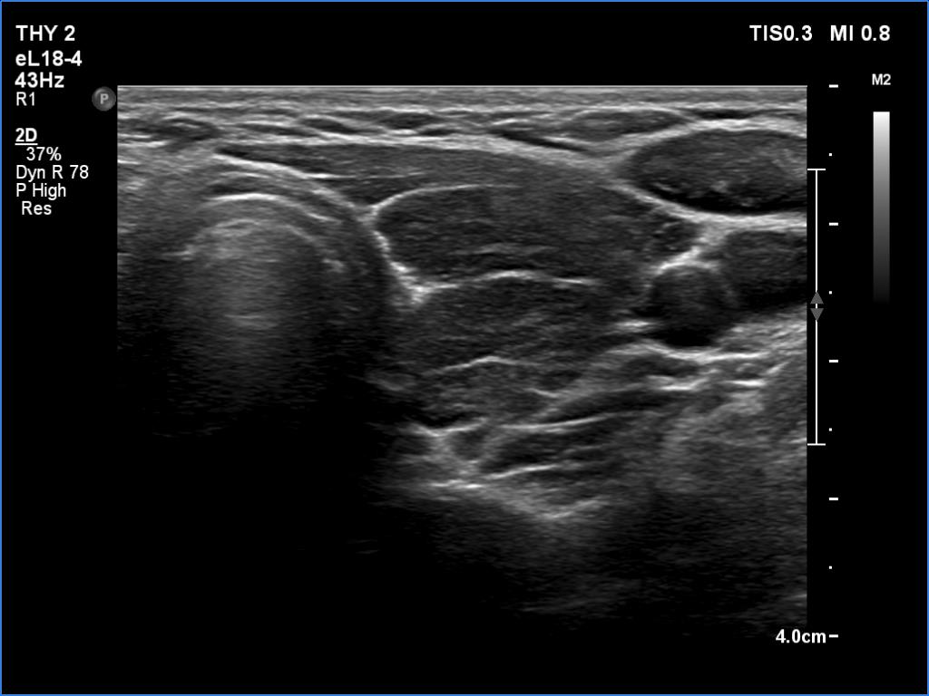
Second examination a year later (second row of images):
Comment. The patient presented a post partum thyroiditis, i.e., the underlying autoimmune thyroiditis became more active. The change in ultrasound pattern is also in line with this. Be aware the rapid change in ultrasound pattern. In other cases, the echogenicity can change in years, rather decades in the case of a hypothyroidism. The structure is expected to show the pattern seen in the first study a year later.Clinical data: Half a year ago, the patient gave birth. She took 100 microgram of medicine a day in the second half of the pregnancy, and after giving birth she returned to the previously used dose of 50 micrograms a day. She gained 8 kg in the last three months and had other symptoms which suggested underdosing.
Palpation: unchanged.
Laboratory tests: TSH 9.88 mIU/L, FT4 7.61 pM/L on daily 50 microgram levothyroxine.
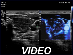
Ultrasonography. In contrast to the previous examination, the entire thyroid became hypoechoic. There were only small islets with less hypoechoic pattern.
Suggestion: to increase the dose of levothyroxine to daily 75 micrograms. Repeat TSH in 6 months.