
|
|
Case 1696
|
|


Clinical data: A 37-yr-old woman was referred for evaluation of a "recurrent papillary cancer" detected on carotid Doppler examination. She was operated on and got radioiodine therapy for a T4 papillary carcer 11 years ago. She was regularly controlled and had no complaints or signs of recurrent tumor.
Palpation: no abnormality.
Laboratory tests: TSH 0.11 mIU/l, FT4 23.3 pM/l, thyroglobulin 0.3 ng/mL) and anti-hTg level below 20 IU/mL on daily 150 microgram levothyroxine.
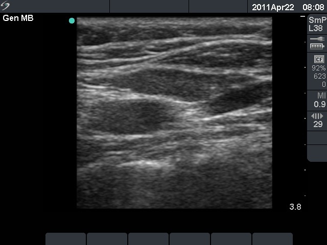
Ultrasonography revealed three hypoechogenic lesions in the left thyroid bed. Two of them were vessels while the third one resembled a lymph node or a thyroid lesion on transverse view. Longitudinal scan disclosed that the lesion in question was in fact a muscle fiber.
Comment. It is very important to perform a thyroid ultrasound examination in two different sections.