
Case 188 (ultrasonographic picture 9)
|
|

|
|
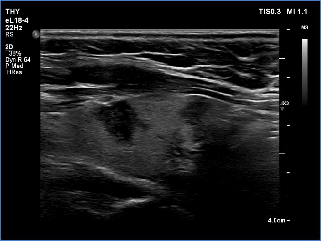
Left lobe, longitudinal scan - set to higher level of harmonization. There are two larger dicrete lesions. Both show nonparallel orientation and irregular borders.