
Case 374 (ultrasonographic picture 1b)
|
|

|
|
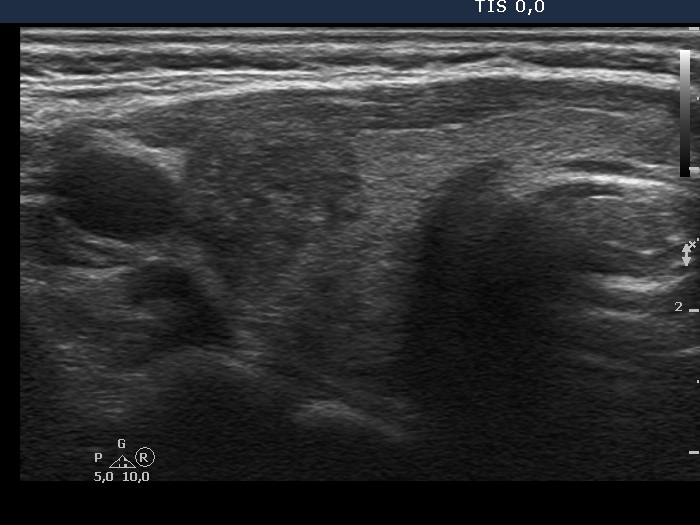
Right lobe, transverse scan. This image illustrates why video is better than still image. You can extract an image from the video at almost any time that not characteristic of the particular thyroid or, as in this case, misleading. Watching the video, it is clear that the hypoechogenic parts have fundamentally blurred borders. But this picture does not show just that. Here, the nodule appears to have a sharp, lobulated margin.