
Case 739
Follow-up investigation two years later (ultrasonographic picture 8)
|
|

|
|
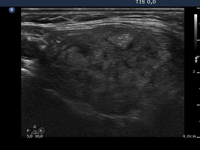
Left lobe, another longitudinal scan. There is a hyperechogenic lesion in the ventral part of the lobe. It contains microcalcifications.