
|
|
Case 922
|
|


Clinical presentation: A 70-year-old woman was referred for evaluation of nodular goiter. We met the patient 10 years ago when FNA was performed from a nodule in the left lobe. At that time, the dimensions of the nodule were 11x9x11 mm, width, depth, length, respectively. The patient had been treated for hypothyroidism for more than 25 years.
Palpation: Both lobes were nodular on palpation.
Laboratory tests: TSH 3.01 mIU/L on daily 62.5 microgram levothyroxine
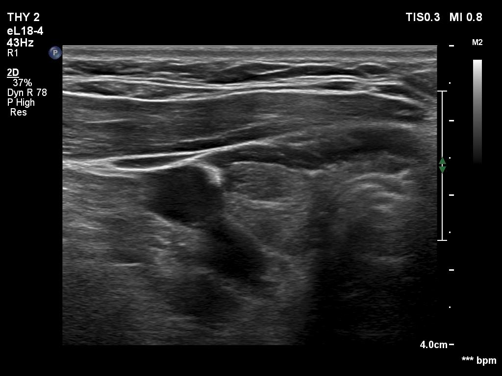
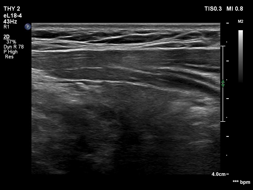
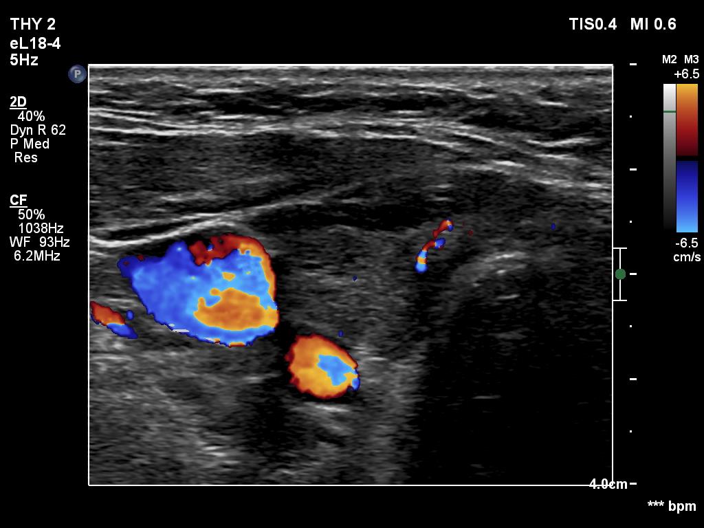
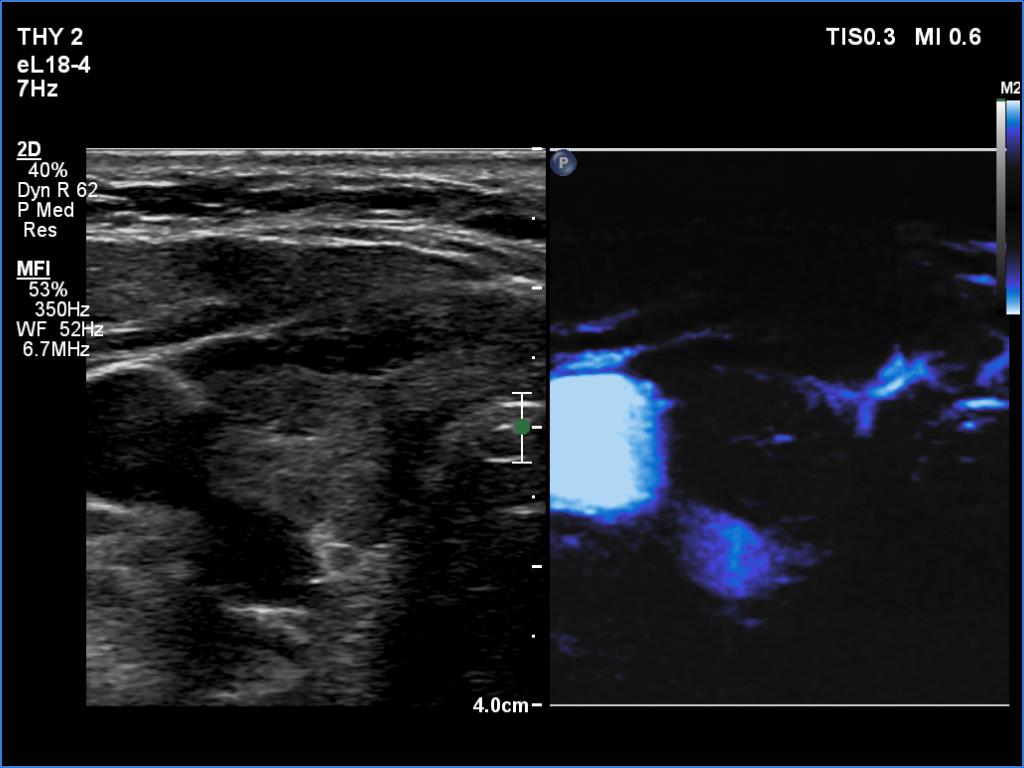
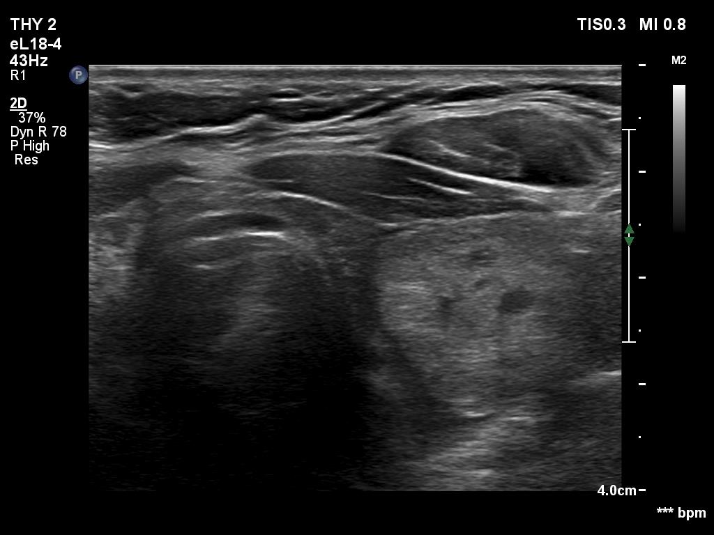
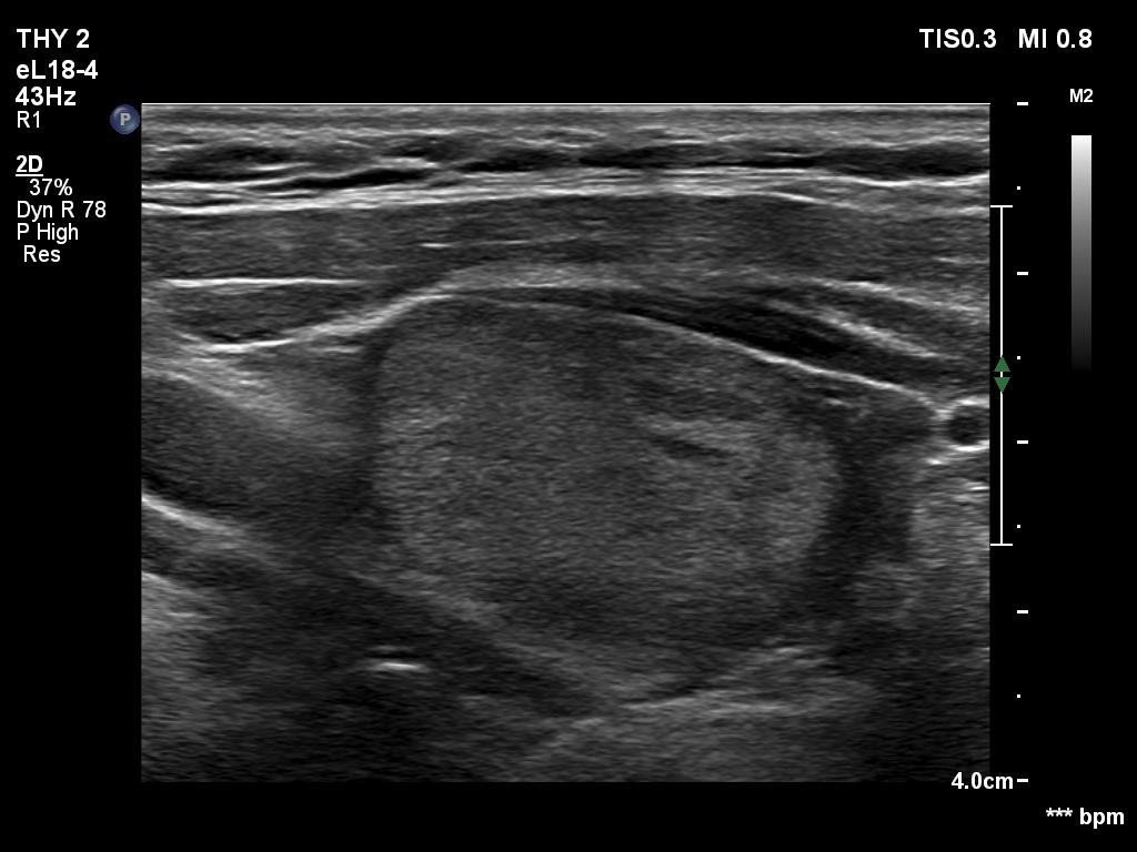
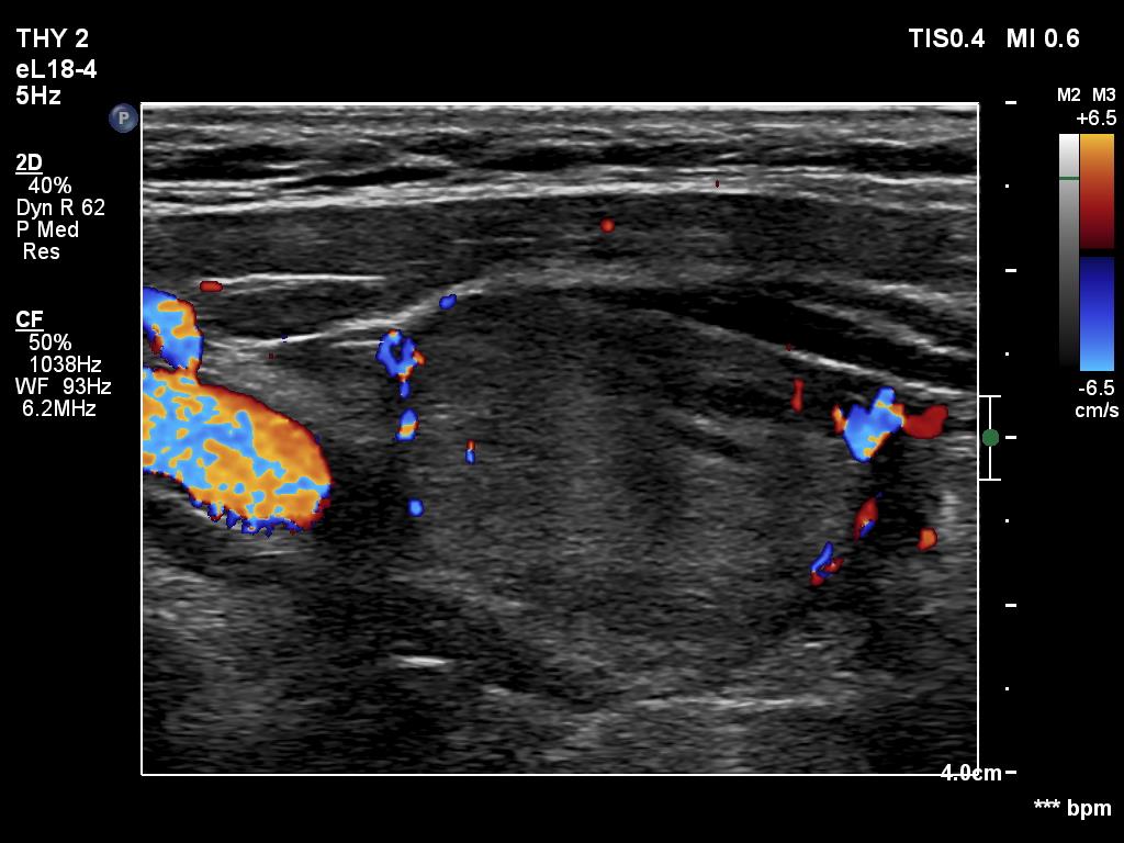
Ultrasonography. The thyroid was hypoechogenic. There was an iso/hyperechoic nodule in both the right and left lobes. The nodule in the left lobe had perinodular vascularity. The dimensions of the nodule in the left lobe were 20x18x31 mm, width, depth, length, respectively. This means that the volume of the nodule has increased almost 8-fold in 10 years.
Cytology of the nodule in the left lobe resulted in benign lesion.