
Case cons100_039 (ultrasonographic picture 7)
|
|

|
|
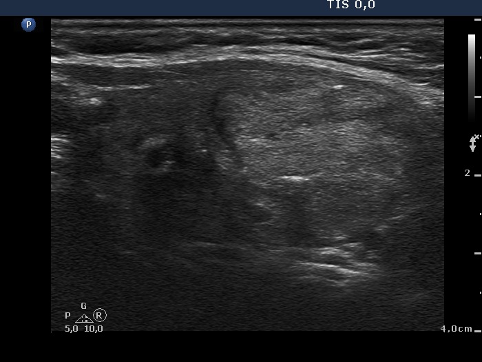
Isthmus and the lower part of the left lobe, longitudinal scan. The lesion is composed of multiple smaller nodules. Note the lobulated margins of the largest nodule.