
|
|
Case cons100_065
|
|


Clinical presentation: A 64-year-old woman was referred for evaluation of a nodular goiter detected on PET CT scan. The latter was performed on follow-up of breast cancer. There was a positive finding in the lower pole of the left lobe.
Palpation: There was a not firm nodule in the lower pole of the left lobe.
Laboratory tests: TSH 1.30 mIU/L, aTPO 1 U/mL.
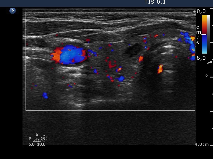
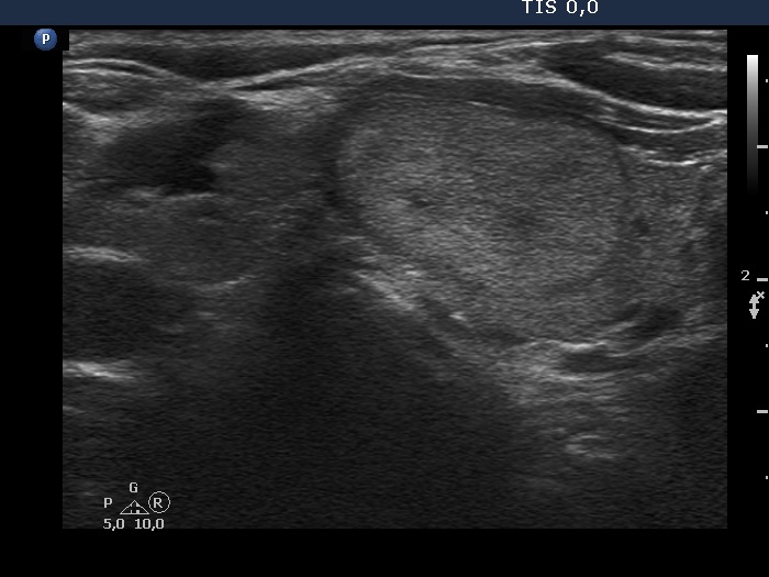
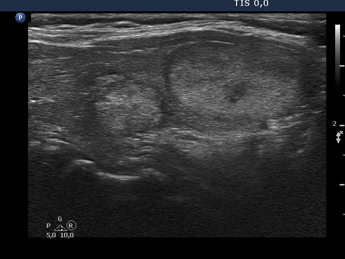
Ultrasonography. The thyroid was moderately hypoechogenic. There were several discrete lesions in the right lobe without any oncological significance. There was a hyperechoic nodule in the lower third of the left lobe, which corresponded to the positive focus on PET CT scan.
Cytology was performed form both from the lesion in the right lobe and from the nodule in the lower part of the left lobe and resulted in benign lesions.
Suggestion: ultrasound in 3 years, TSH in a year.
Comment. The basic pattern of the thyroid raises the suspicion of an autoimmune thyroiditis despite the normal aTPO level. In such cases we routinely propose yearly TSH check.