
|
|
Thyroid cancers - case 586
|

|


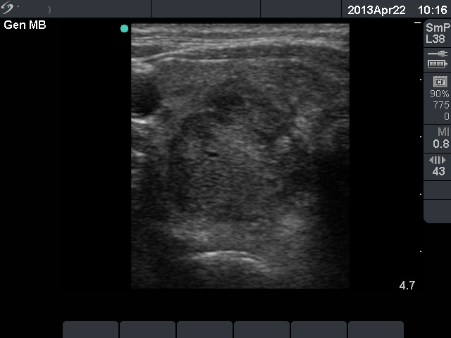
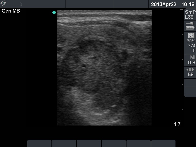
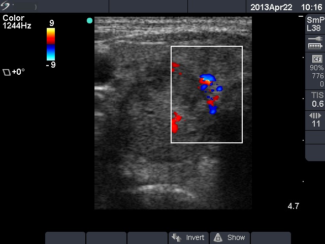
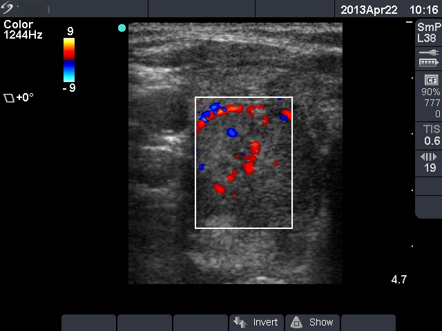
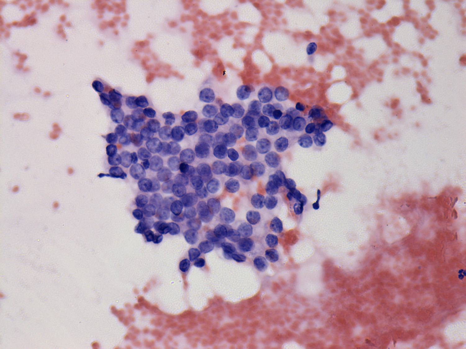
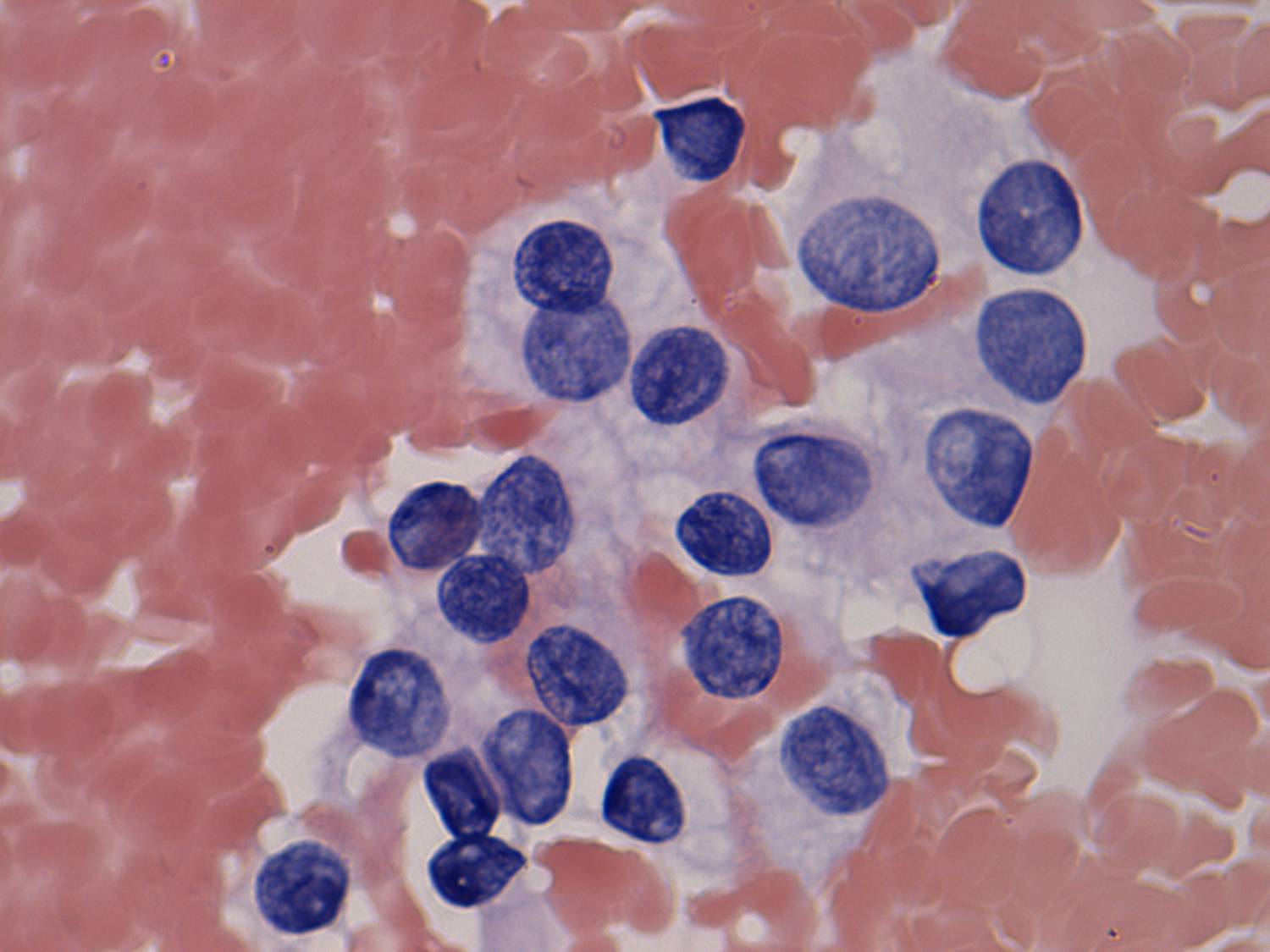
Clinical presentation: A 69-year-old woman was referred for aspiaration cytology because a nodule was detected on carotid Doppler examination. The patient had been operated on clear cell renal cancer for 6 years and was regularly checked up. No clinical or radiological sign of a recurrence had been detected so far.
Palpation: The right lobe had a firm nodule.
Laboratory test: TSH 1.99 mIU/L.
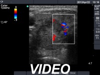
Ultrasonography. The thyroid was echonormal. There was a large moderately hypoechoic nodule in the lower-dorsal part of the right lobe. The lesion presented with irregular borders and had both intranodular and perinodular vascularity. A small cystic lesion was found in the left lobe.
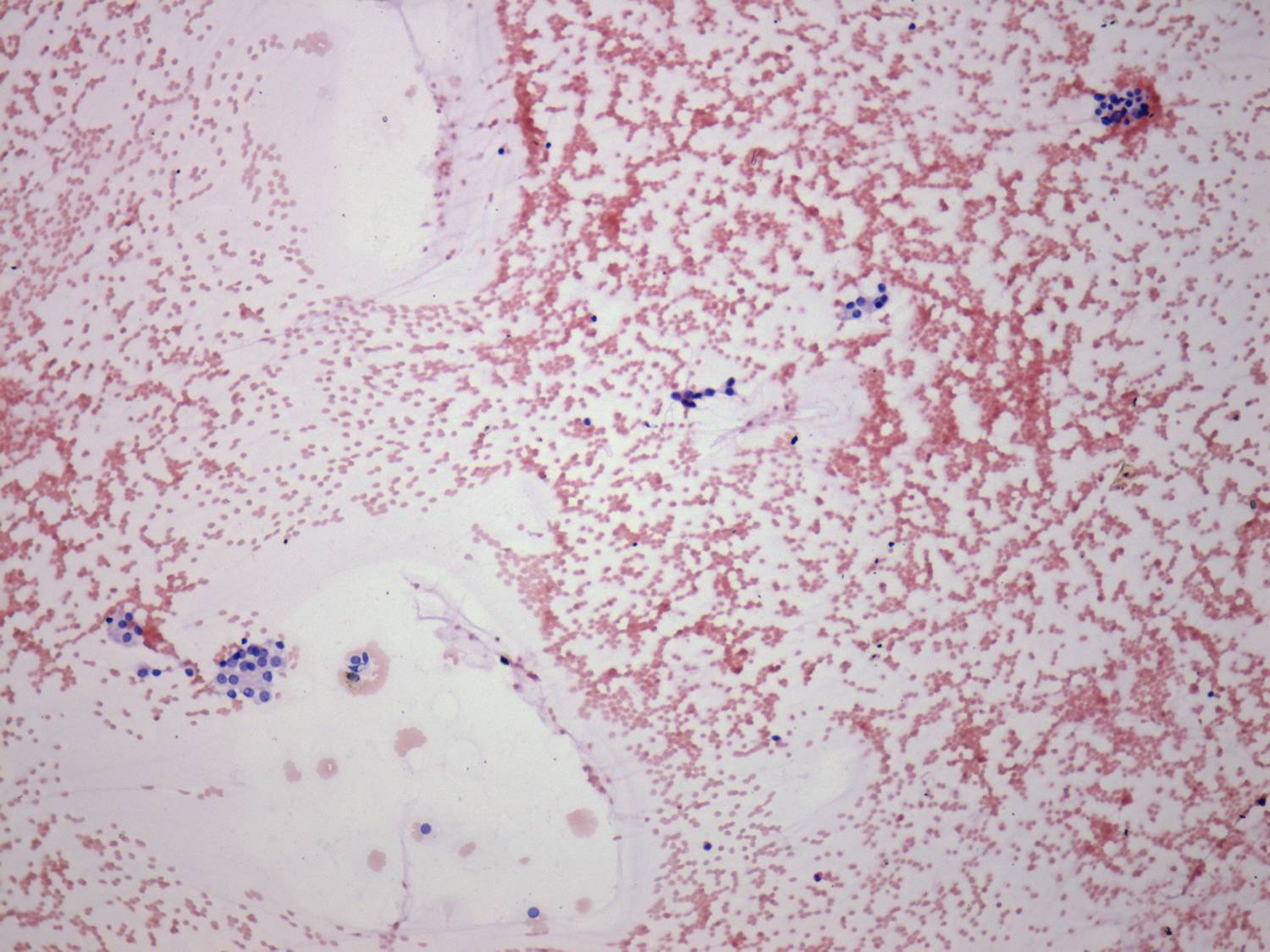
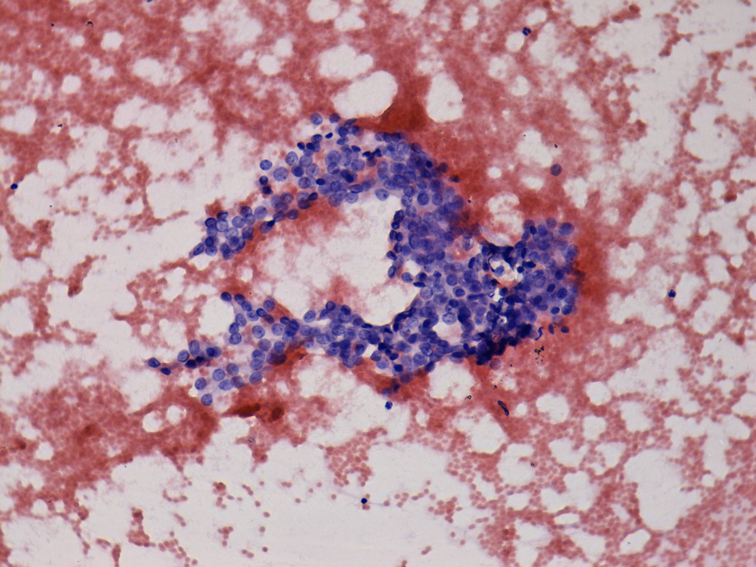
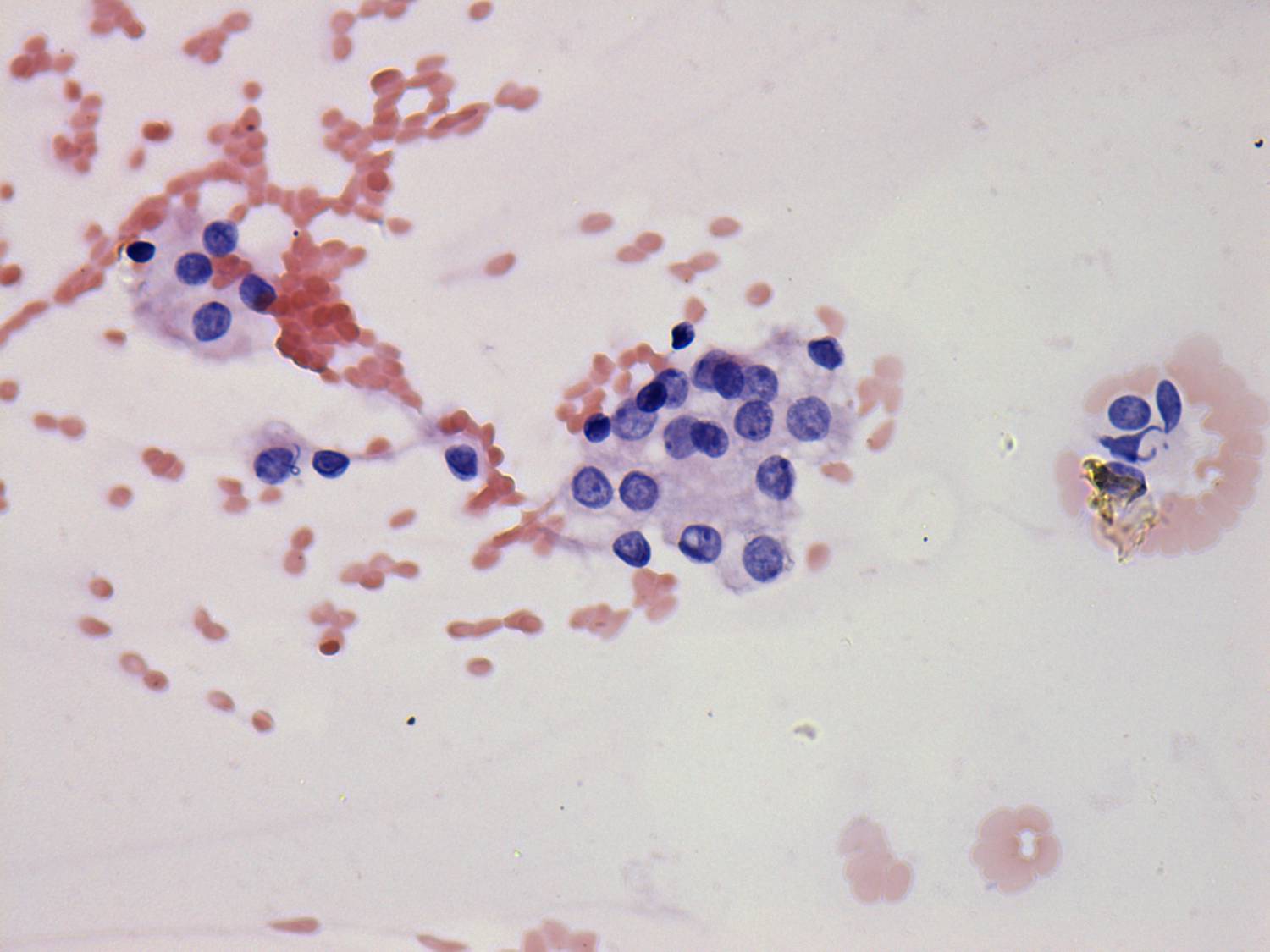
Cytological picture. There was no colloid in the background. Epithelial cells were mostly in non-specific cell groups. They showed anisonucleosis and had prominent nucleoli. The cytoplasm of the cells has vacuoles. On the cytological presentation, a benign nodular goiter, an oxyphilic lesion and a metastatic clear cell renal cancer could be considered.
Wash-out thyroglobulin resulted in 1.01 ng/dL.
Our final diagnosis was metastasis of clear cell renal cancer.
On further evaluation, no metastatic foci were found, and the oncological team suggested surgery. Histopathology disclosed metastasis of clear cell renal cancer.
Comment. Neither the ultrasound nor the cytological presentation was suspicious of secondary cancer. The patient' history was the pivotal as in most cases of secondary thyroid tumors.


