
|
|
Study on 100 consecutive patients with thyroid nodule - case 005
|
|


Clinical presentation: A 51-year-old man was referred for evaluation of a longstanding nodule. On lung screening, a substernal goiter was detected. The patient had no complaints.
Palpation: The right lobe was enlarged and nodular on palpation.
Functional state: euthyroidism (TSH 0.12 mIU/L, FT4 14.6 pM/L, FT3 4.79 pM/L).
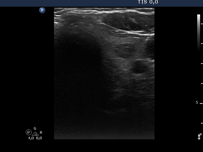
Ultrasonography. The right lobe was greatly enlarged and had numerous nodules. The lower pole could not be visualized during swallowing. The left lobe had a small, moderately hypoechoic nodule.
Cytology was performed from one of the nodules in the right lobe and from the lesion in the left lobe. Both resulted in benign colloid goiter.
Neck and upper mediastinal CT scan were performed which showed that the right lobe spread almost 11 cm below the level of the clavicle.
Right lobectomy was performed. The removal of the right thyroid gland could not be performed from a low incision, the surgery had to extended with sternotomy. Histopathology disclosed benign hyperplastic nodules.
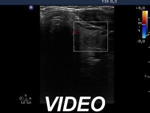
Comments. It is worth viewing the video to learn how to define the degree of substernal spread on ultrasound.