
Study on 100 consecutive patients with thyroid nodule - case 016 (ultrasonographic picture 2)

|

|

|
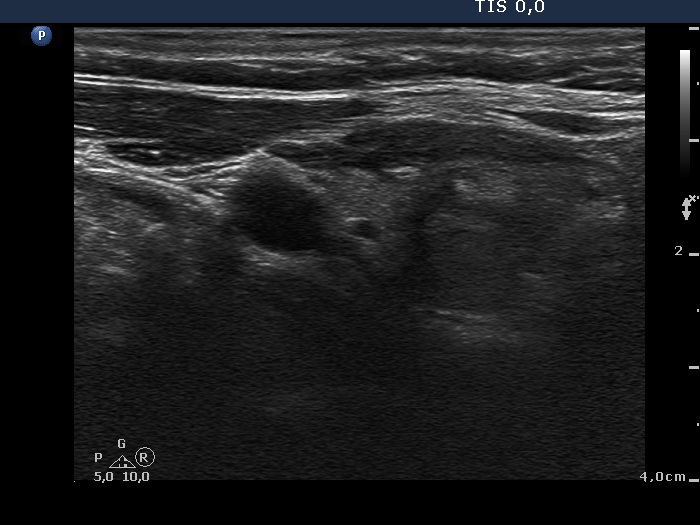
Right lobe, longitudinal scan. The upper part of the lobe is echonormal while the lower two-third is somewhat hypoechogenic and contains a ventral hyperechogenic lesion and a dorsal minimally hypoechogenic area. The pattern resembles Hashimoto's thyroiditis.


