
Study on 100 consecutive patients with thyroid nodule - case 021 (ultrasonographic picture 5)
|
|

|
|
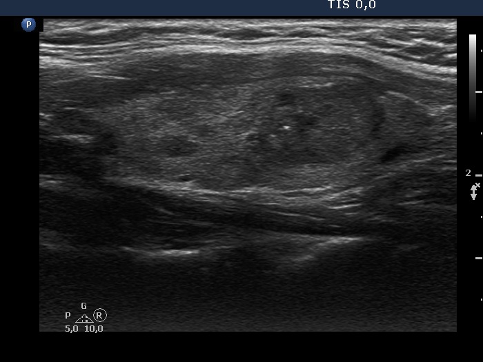
Left lobe, longitudinal scan. It would be hard to interpret the brightest echogenic granule other than a microcalcification.