
|
|
Study on 100 consecutive patients with thyroid nodule - case 023
|
|


One year before the present examination (first row of images)
Clinical presentation: A 55-year-old man was referred for examination of a lump in the neck which has developed for 2 weeks.
Palpation: an elastic nodule in the right lobe.
Functional state: euthyroidism (TSH 0.97 mIU/L).
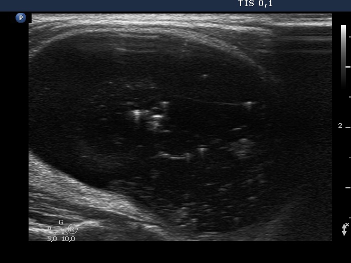
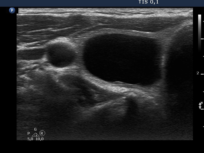
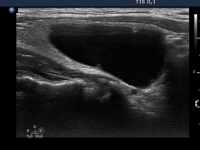
Ultrasonography. The thyroid was echonormal. A large nodule occupied almost the entire right lobe. This was an almost completely cyst with a tiny echonormal solid part at the ventral wall. The nodule had numerous large comet-tail artifacts and showed no vascularity. The dimensions of the nodule were 24x27x48 mm - 16.3 mL.
Only 2.5 mL brown fluid could be aspirated from the cyst. Cytology resulted in benign cystic degeneration.
Suggestion: repeat examination in 4 months.
Present examination years after the first examination (third row of images)
Clinical presentation: The patient had no complaints. He noticed a decrease in nodule size.
Palpation: an elastic nodule in the right lobe.
Functional state: euthyroidism (TSH 3.04 mIU/L).
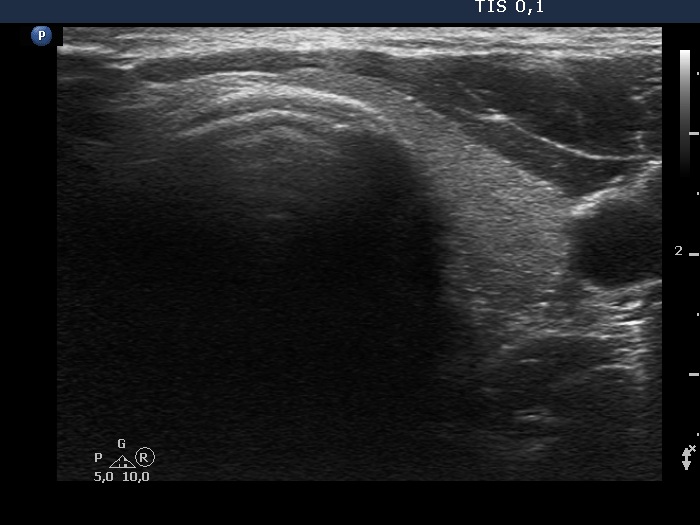
Ultrasonography. Except for the size of the nodule, the pattern remained the same. The dimensions of the nodule were 23x15x33 mm - 5.95 mL.
Suggestion: ultrasound in two years.
Comments.
-
At the first examination, the nodule compressed the vessels running outside the nodule while on follow-up this compression was released, therefore the perinodular vascularity became visible.
-
The nonparallel orientation became parallel.
- If we can remove only small proportion of cystic content, we usually offer a follow up within 6 months. In around third of cases, the thick content flows out and we can aspirate the cystic part.