
Study on 100 consecutive patients with thyroid nodule - case 024 (ultrasonographic picture 8)

|

|

|
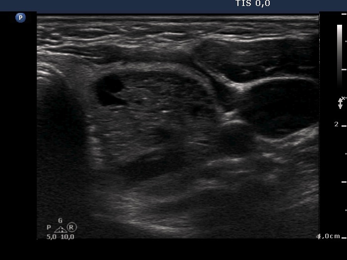
Lower part of the left lobe, transverse scan. This portion of the nodule is solid and presents numerous hyperechogenic figures, mostly related to tiny, ventral cystic areas. These figures are caused by back wall posterior enhancement.


