
|
|
Study on 100 consecutive patients with thyroid nodule - case 027
|

|


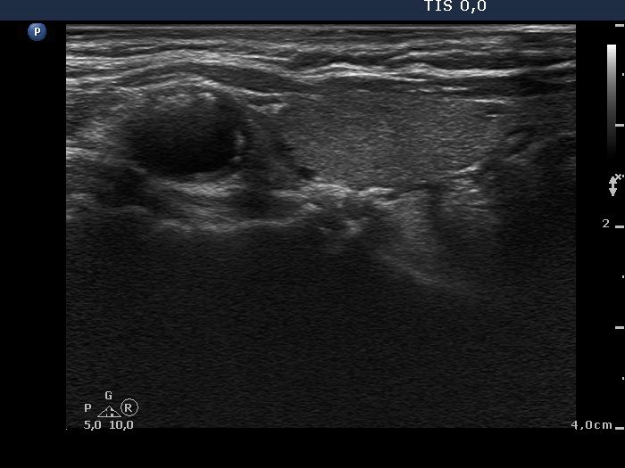
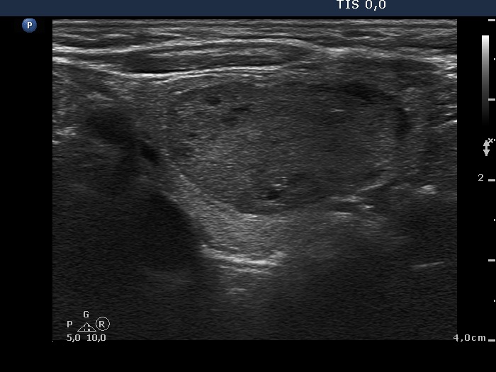
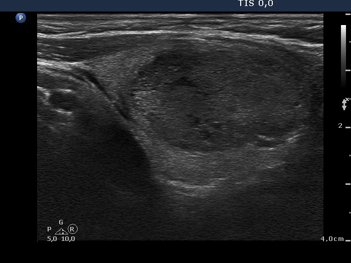



Clinical presentation: An 81-year-old woman was referred for evaluation of a nodular goiter discovered by her family physician.
Palpation: a moderately firm nodule in the lower part of the right lobe.
Functional state: euthyroidism (TSH 2.09 mIU/L).
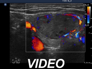
Ultrasonography. The thyroid was minimally hypoechogenic. There was a discrete lesion in the upper part of the right lobe. It was equivocal whether this echo abnormality was a nodule or not. A relatively large hypoechogenic, inhomogeneous nodule was in the lower half of the right lobe. This nodule did not present halo sign but did a perinodular blood flow.
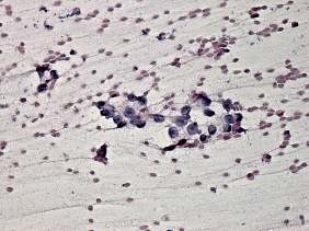
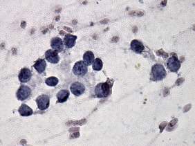
Aspiration cytology of the large nodule resulted in microfollicular proliferation without significant atypia. Cytological diagnosis was follicular tumor.
Considering the echo pattern, we gave a final diagnosis of follicular tumor with less than 2% risk of malignancy.
We advised yearly follow-up instead of surgery.
Comments.
-
In our practice with more than 5,000 cyto-histological comparisons, the risk of carcinoma in a patient with cytological result of follicular tumor without significant atypia is around 2-3%. If we take the ultrasound presentation into account, the risk may be less or more, the risk ranging from 0.5% to 10%.
-
Considering the small risk of follicular carcinoma and the age of the patient, the risk of surgery seemed to be significantly higher than the risk of avoidance of surgery.


