
|
|
Study on 100 consecutive patients with thyroid nodule - case 030
|
|


Clinical presentation: A 47-year-old woman came to a follow-up visit. We met her 12 years ago when 16 mL yellow fluid was aspirated form a nodule in the right lobe with the dimensions of 38x28x47 mm. There was a minimally hypoechoic lesion in the left lobe with the dimensions of 6x5x6 mm. Until recently, the cyst has not relapsed, but 2 weeks before the examination the patient noticed the appearance of the lump in the right lobe.
Palpation: a firm nodule in the right lobe.
Functional state: euthyroidism (TSH 1.73 mIU/L).
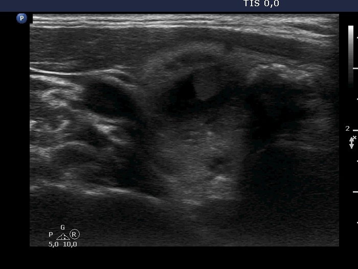
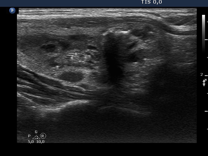
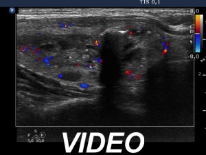
Ultrasonography. The thyroid was echonormal. There were two nodules in the right lobe, a dominantly cystic one, and a lesion in the lower third which had macrocalcifications. There was a minimally hypoechoic lesion in the left lobe which showed halo sign and perinodular vascularity. Compared with the first examination, the nodule in the left lobe showed a substantial increase, this time the dimensions were 15x14x17 mm.
6 mL brown fluid was removed from the cystic nodule and we performed FNA from the left lesion, too. Cytology resulted in benign cystic lesion and follicular proliferation, right and left nodule, respectively.
We suggested surgery with relative indication based on the ultrasound and cytological appearance of the nodule in the left lobe. The patient wished not to be operated. We agreed that she will continue with regular ultrasound check and if the nodule in the left lobe would further increase, she will accept my proposal.
Comments.
-
The echogenicity of the solid part of a cystic nodule is significantly influenced by the location. In this case, the solid part was echonormal dorsal to the cystic content while moderately hypoechoic in the medial part. The difference in echogenicity is explained by the echo amplification dorsal to the fluid.
-
The echogenic figures are better to judge before aspiration. These were clearly back wall figures and comet-tail artifacts before the aspiration.
-
The ultrasound presentation of the left lobe is almost diagnostic of follicular tumor, a solitary, homogeneous lesion presenting halo and perinodular blood flow has more than 90% risk being a follicular tumor.