
Study on 100 consecutive patients with thyroid nodule - case 030 (ultrasonographic picture 3)
|
|

|
|
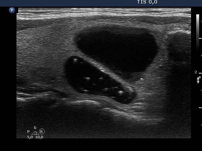
Right lobe, another longitudinal view. Typical comet-tail artifacts can be seen. Note the bright granule in the lower part (right in the image). This is located dorsal to a tiny cystic area, therefore it should not be regarded as a microcalcification.