
Study on 100 consecutive patients with thyroid nodule - case 057 (ultrasonographic picture 8)

|

|

|
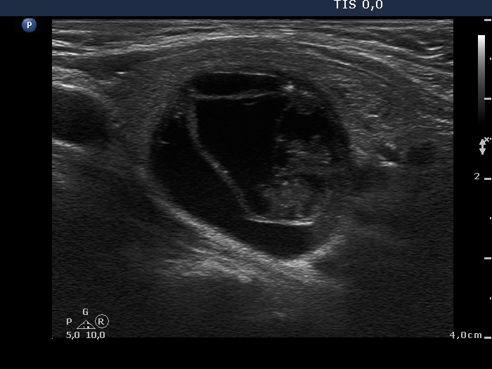
Left lobe, longitudinal scan. This nodule has minimally hypoechogenic solid areas with both echogenic lines and granules. The latter correspond to back wall figures caused by posterior enhancement.


