
Study on 100 consecutive patients with thyroid nodule - case 063 (ultrasonographic picture 6)

|

|

|
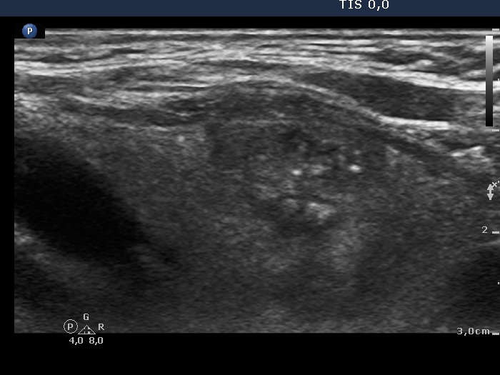
Left lobe, longitudinal view, enlargement. The two brightest granules probably correspond to microcalcifications. Note the lobulated margins.


