
Consecutively operated patients with autoimmune thyroid disease - case 16 (50) (ultrasonographic picture 9)
|
|

|
|
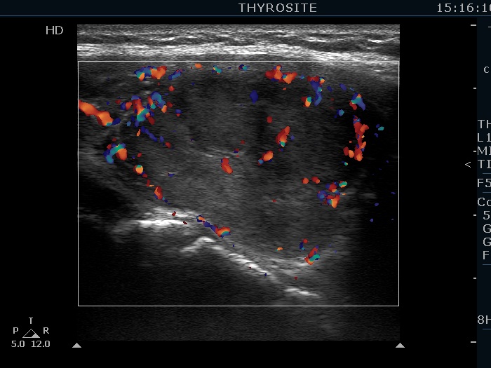
Left lobe, longitudinal view, power Doppler mode. The nodule shows sign of perinodular vascularity, as well.