
Discrete lesion or nodule in Hashimoto's thyroiditis - case 10 (95)
Follow-up investigation 38 months after the first visit (ultrasonographic picture 7)
|
|

|
|
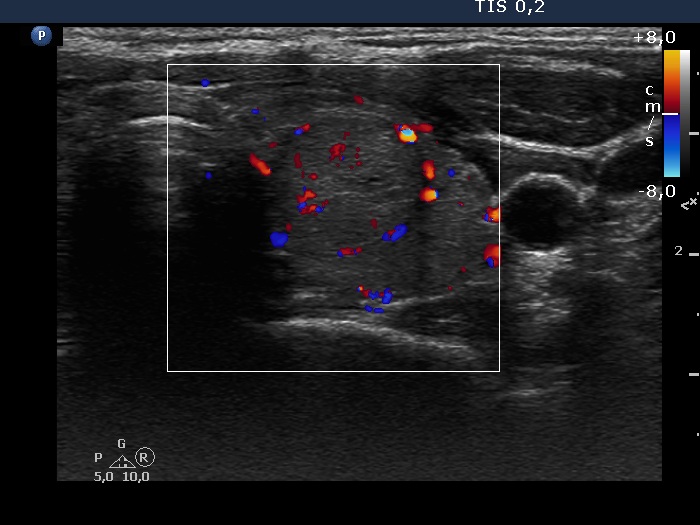
Left lobe, transverse scan, color Doppler mode. The lesion presents signs of perinodular blood flow.